. 2007 Nov 15;2(5):316-27.
doi: 10.1016/j.chom.2007.10.002.

Heparan sulfate proteoglycans provide a signal to Plasmodium sporozoites to stop migrating and productively invade host cells

Affiliations

Heparan sulfate proteoglycans provide a signal to Plasmodium sporozoites to stop migrating and productively invade host cells

Alida Coppi et al. Cell Host Microbe. .

Abstract

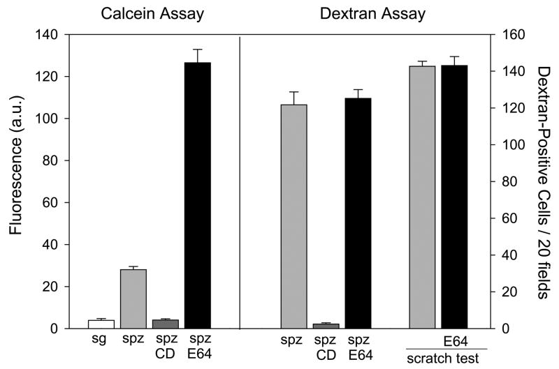
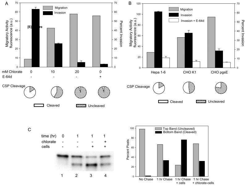
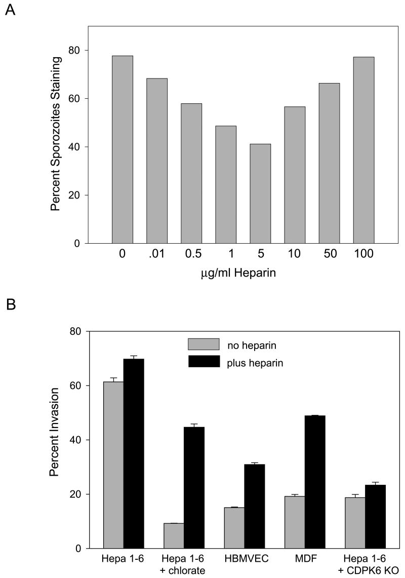
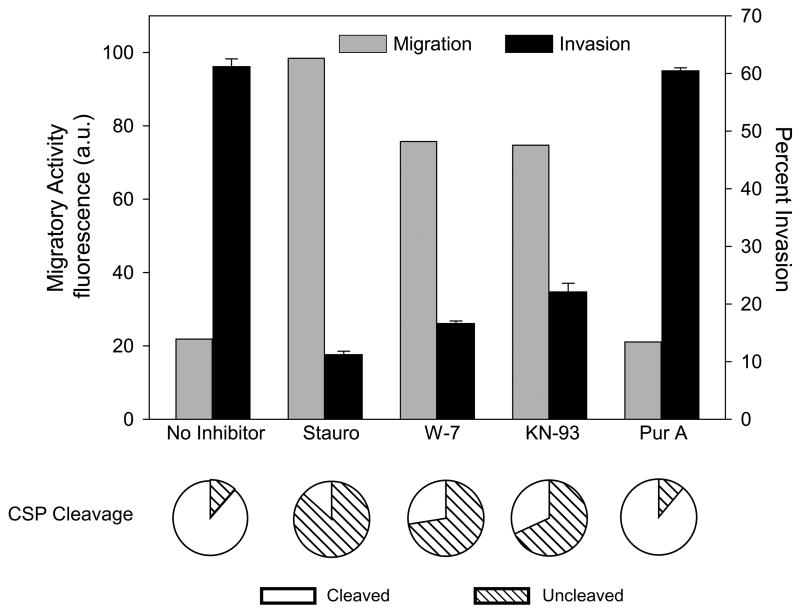
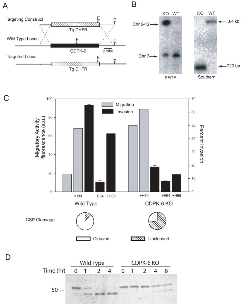
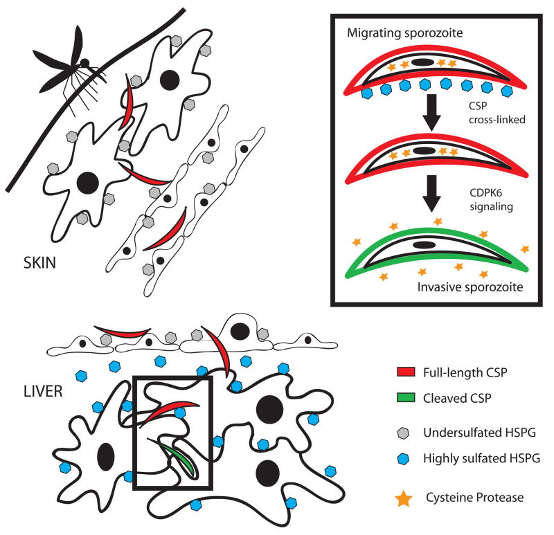
Malaria infection is initiated when Anopheles mosquitoes inject Plasmodium sporozoites into the skin. Sporozoites subsequently reach the liver, invading and developing within hepatocytes. Sporozoites contact and traverse many cell types as they migrate from skin to liver; however, the mechanism by which they switch from a migratory mode to an invasive mode is unclear. Here, we show that sporozoites of the rodent malaria parasite Plasmodium berghei use the sulfation level of host heparan sulfate proteoglycans (HSPGs) to navigate within the mammalian host. Sporozoites migrate through cells expressing low-sulfated HSPGs, such as those in skin and endothelium, while highly sulfated HSPGs of hepatocytes activate sporozoites for invasion. A calcium-dependent protein kinase is critical for the switch to an invasive phenotype, a process accompanied by proteolytic cleavage of the sporozoite's major surface protein. These findings explain how sporozoites retain their infectivity for an organ that is far from their site of entry.


Figures

Figure 1
Figure 1. A new sporozoite migration assay
Sporozoites pre-incubated ± E-64d (E64) were added to Hepa 1–6 cells and migratory activity was measured using two different assays. On the left is the calcein assay in which sporozoites were incubated for 1 hr with Hepa 1–6 cells pre-loaded with calcein and the amount of fluorescent calcein released into the medium was measured using a fluorimeter. Fluorescence is expressed as arbitrary units (a.u.). On the right is the dextran assay in which sporozoites were added to Hepa 1–6 cells in the presence of dextran-FITC and the number of fluorescent cells per 20 fields is shown. Controls include salivary glands from uninfected mosquitoes (sg) and sporozoites (spz) treated with cytochalasin D (CD). As a control for the effect of E-64d on wound healing, Hepa 1–6 cells ± E-64d were wounded by scratching with a razor blade in the presence of dextran-FITC, allowed to heal for 1 hr and the number of FITC-positive cells in 100 fields were counted (scratch test). Shown are means ± SD of triplicates.
Figure 2
Figure 2. Highly sulfated HSPGs arrest sporozoite migration and trigger invasion and CSP processing
(A) Effect of chlorate treatment on migration, invasion and CSP processing. Hepa 1–6 cells were grown in the presence of the indicated amounts of chlorate, washed and P. berghei sporozoites preincubated ± E-64d were added for 1 hr (migration and invasion assays) or 3 minutes (CSP cleavage assay). To measure migration (grey bars), chlorate-treated Hepa 1–6 cells were preloaded with calcein green, sporozoites were added and the fluorescence released into the supernatant was measured. Shown are the means of duplicates which did not vary more than +/− 5%. Invasion (black bars) was quantified by fixing, staining and counting the numbers of intracellular and extracellular sporozoites. Shown are means ± SD of triplicates. Proteolytic cleavage of CSP (pie charts) was quantified by adding GFP-expressing sporozoites to cells for 3 min, fixing and staining with antiserum specific for full-length CSP. 100 sporozoites per coverslip were counted and the pie charts show the percentage of sporozoites with cleaved (white) or full-length (hatched) CSP. Shown are the means of triplicates which did not vary more than +/− 5%. All experiments were performed at least 3 times and shown are representative experiments. (B) A CHO cell mutant in HSPG sulfation enhances sporozoite migratory activity and inhibits invasion and CSP cleavage. Sporozoites were incubated with the indicated cell type and migration (grey bars), invasion in the absence (black bars) or presence (white bars) of E-64d, and CSP cleavage (pie charts) were quantified as described above. For the invasion assay, shown are means ± SD of triplicates. Migratory activity is measured as fluorescence a.u. and shown are the means of duplicates which did not vary more than +/− 5%. Pie charts show the means of triplicates which did not vary more than +/− 5%. All experiments were performed 3 times and shown is a representative experiment. (C) Quantification of CSP cleavage induced by highly sulfated HSPGs. Sporozoites were metabolically labeled with 35[SO4]-Cys/Met and kept on ice (lane 1) or chased for 1 hr (lanes 2–4). They were then spun onto coverslips without cells (lanes 1 & 2) or with cells grown in the absence (lane 3) or presence (lane 4) of chlorate and brought to 37°C for 3 min. Cells and sporozoites were then lysed, CSP was immunoprecipitated and analyzed by SDS-PAGE and autoradiography. Band intensity was quantified by densitometry and is displayed graphically on the right; grey bars represent uncleaved CSP and black bars represent cleaved CSP.
Figure 3
Figure 3. Sporozoite contact with endothelial cells and dermal fibroblasts leads to increased migratory activity and decreased productive invasion and CSP cleavage
(A) Sporozoites were incubated with Hepa 1–6 cells, dermal fibroblasts (MDF) or an endothelial cell line (HBMVEC) and migration (grey bars), invasion in the absence (black bars) or presence (white bars) of E-64d and CSP cleavage (pie charts) were quantified as previously described. For the invasion assay, shown are means ± SD of triplicates. Migratory activity is measured as fluorescence a.u. and shown are the means of duplicates which did not vary more than +/− 5%. Pie charts show the means of triplicates which did not vary more than +/− 5%. All experiments were performed 3 times and shown are representative experiments. (B) Development of EEFs in dermal fibroblasts and endothelial cells. Sporozoites were added to the indicated cell line and 44 hr later cells were fixed, stained and EEFs were counted. Shown are means ± SD of triplicates. This experiment was performed 3 times and shown is a representative experiment.
Figure 4
Figure 4. Soluble heparin induces CSP cleavage and enhances sporozoite invasion of nonpermissive cells
(A) CSP cleavage after a 5 min incubation with the indicated concentrations of heparin was quantified by fixing and staining sporozoites with antisera specific for full-length CSP. 200 sporozoites per well were counted and shown is the percentage of sporozoites staining. This experiment was repeated 3 times with identical results. (B) Sporozoite invasion of the indicated cell line was quantified by adding wild type or CDPK-6 mutant (CDPK6 KO, see below) sporozoites preincubated for 5 min with medium alone (grey bars) or medium containing 5 μg/ml heparin (black bars) to Hepa 1–6 cells grown in the absence or presence of chlorate, HBMVEC or MDF cells. After 1 hr, cells were fixed, stained and the numbers of intracellular and extracellular sporozoites were counted. Shown are means ± SD of triplicates. This experiment was repeated twice with identical results.
Figure 5
Figure 5. Kinase inhibitors enhance sporozoite migration, inhibit invasion and decrease CSP cleavage
P. berghei sporozoites preincubated with the indicated inhibitors were added to Hepa 1–6 cells for 1 hr (migration and invasion assays) or 3 min (CSP cleavage assay). Migration (grey bars) invasion (black bars) and proteolytic cleavage of CSP (pie charts) were quantified as previously outlined. For the invasion assay shown are means ± SD of triplicates. Migration is measured as fluorescence a.u. and shown are the means of duplicates which did not vary more than +/− 5%. Pie charts show the means of triplicates which did not vary more than +/− 5%. All experiments were performed 3 times and shown are representative experiments. Stauro, staurosporine; Pur A, purvalanol A.
Figure 6
Figure 6. Construction and phenotypic characterization of CDPK-6 mutant sporozoites
(A) Strategy used to target the CDPK-6 locus (black box). The targeting construct contained the selectable marker cassette (T. gondii DHFR-TS with upstream and downstream control elements of P. berghei DHFR-TS; hatched box) flanked by the 5' UTR of CDPK-6 (thick grey line) and the 3'UTR of CDPK-6 (thick white line) to target recombination. EcoRV sites are labeled E. (B) Confirmation of genotype. Transfected clones were checked by PFGE using a probe specific for the 3'UTR of P. berghei DHFR-TS which detects the wild type DHFR-TS locus on chromosome 7 and the altered CDPK-6 locus in the transgenic parasites consistent with its location on chromosome 9. Clones were also checked by Southern blot after digestion of genomic DNA with EcoRV. The probe specific for the 3'UTR of CDPK-6 (black line in A) detects diagnostic fragments of 3.4 kb in wild type and 725 bp in transgenic parasites. (C) Wild type or CDPK-6 mutant P. berghei sporozoites were preincubated in medium alone, the PKA inhibitor H-89 or E-64d as indicated, added to Hepa 1–6 cells and migration (grey bars), invasion (black bars) or CSP cleavage (pie charts) were measured as previously outlined. For the invasion assay, shown are means ± SD of triplicates. Migration is measured as fluorescence a.u. and shown are the means of duplicates which did not vary more than +/− 5%. Pie charts show the means of triplicates which did not vary more than +/− 5%. All experiments were performed twice and shown are representative experiments. (D) Wild type or CDPK-6 mutant P. berghei sporozoites were metabolically labeled with 35[SO4]-Cys/Met and kept on ice (time 0) or chased for the indicated times. Sporozoites were then lysed, CSP was immunoprecipitated and analyzed by SDS-PAGE and autoradiography.
Figure 7
Figure 7. Model of sporozoite activation for invasion by highly sulfated HSPGs
Migratory sporozoites (red) are injected into the dermis by an infected mosquito where they encounter cells expressing undersulfated HSPGs (grey hexagons). They traverse endothelial cells, also expressing undersulfated HSPGs and enter the bloodstream. In the liver they cross the sinusoidal barrier and encounter the highly sulfated HSPGs found in the loose basement membrane of the liver (space of Disse) and on hepatocytes (blue hexagons) and become activated for productive invasion (green sporozoites). The inset shows some of the specific steps involved in sporozoite activation, namely crosslinking of CSP by highly sulfated HSPGs which results in a CDPK-6 dependent signaling pathway associated with the secretion of a parasite cysteine protease (stars) that proteolytically processes surface CSP.

Comment in

References

    1. Amino R, Thiberge S, Martin B, Celli S, Shorte S, Frischknecht F, Menard R. Quantitative imaging of Plasmodium transmission from mosquito to mammal. Nat Med. 2006;12:220–224. - PubMed
    1. Bame KJ, Esko JD. Undersulfated heparan sulfate in a Chinese Hamster Ovary cell mutant defective in heparan sulfate N-sulfotransferase. J Biol Chem. 1989;264:8059–8065. - PubMed
    1. Bhanot P, Schauer K, Coppens I, Nussenzweig V. A surface phospholipase is involved in the migration of Plasmodium sporozoites through cells. J Biol Chem. 2005;280:6752–6760. - PubMed
    1. Billker O, Dechamps S, Tewari R, Wenig G, Franke-Fayard B, Brinkmann V. Calcium and a calcium-dependent protein kinase regulate gamete formation and mosquito transmission in a malaria parasite. Cell. 2004;117:503–514. - PubMed
    1. Carruthers VB, Sibley LD. Mobilization of intracellular calcium stimulates microneme discharge in Toxoplasma gondii. Mol Microbiol. 1999;31:421–428. - PubMed

Publication types

Substances