. 2008 Jan 25;283(4):2407-17.
doi: 10.1074/jbc.M703612200. Epub 2007 Nov 15.

Inhibition of Raf-1 alters multiple downstream pathways to induce pancreatic beta-cell apoptosis

Affiliations

Inhibition of Raf-1 alters multiple downstream pathways to induce pancreatic beta-cell apoptosis

Emilyn U Alejandro et al. J Biol Chem. .

Abstract

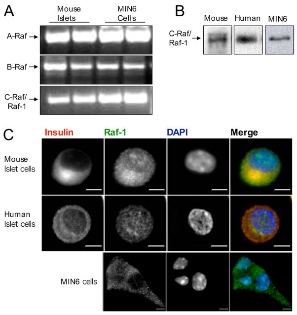
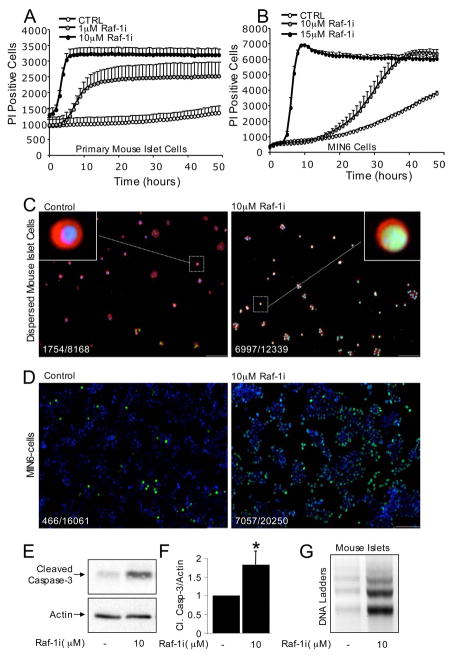
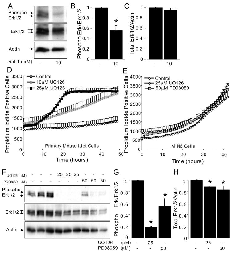
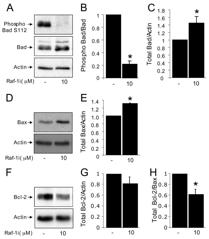
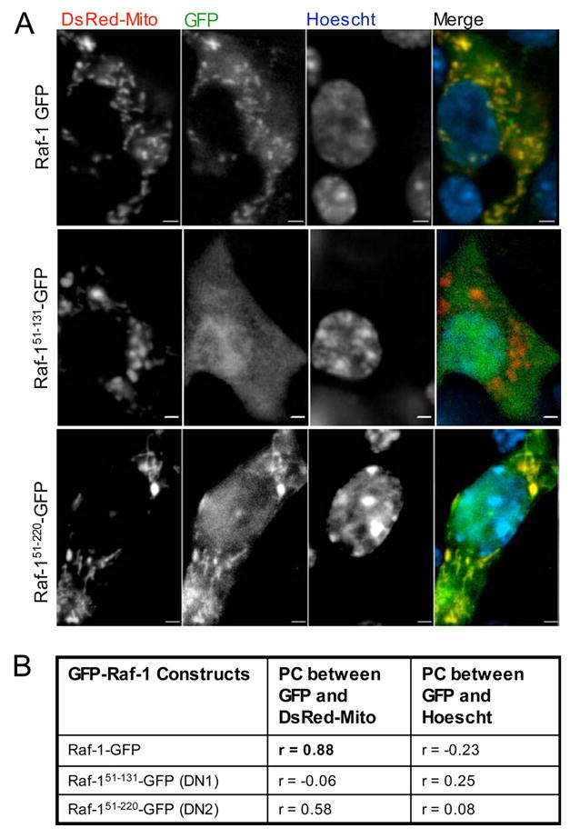
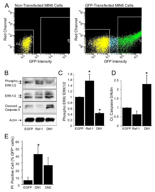
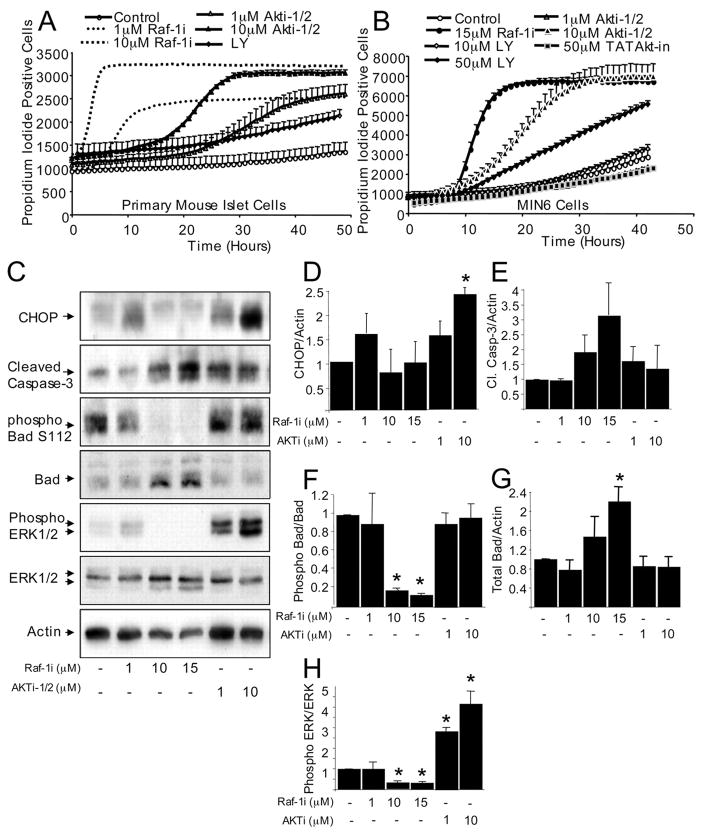
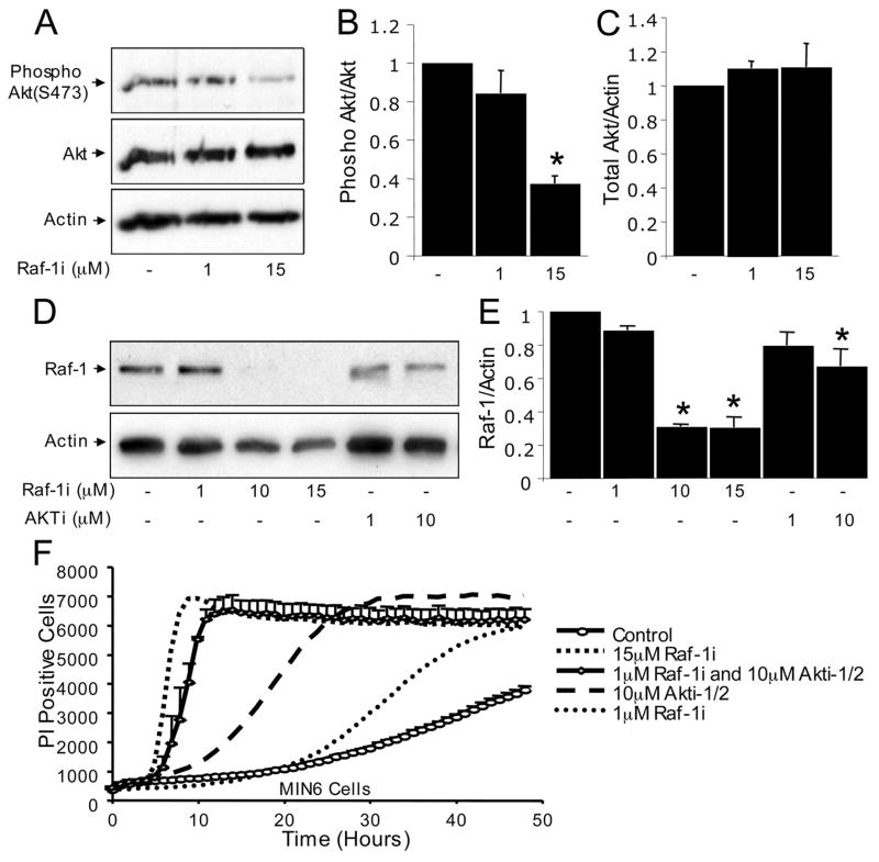
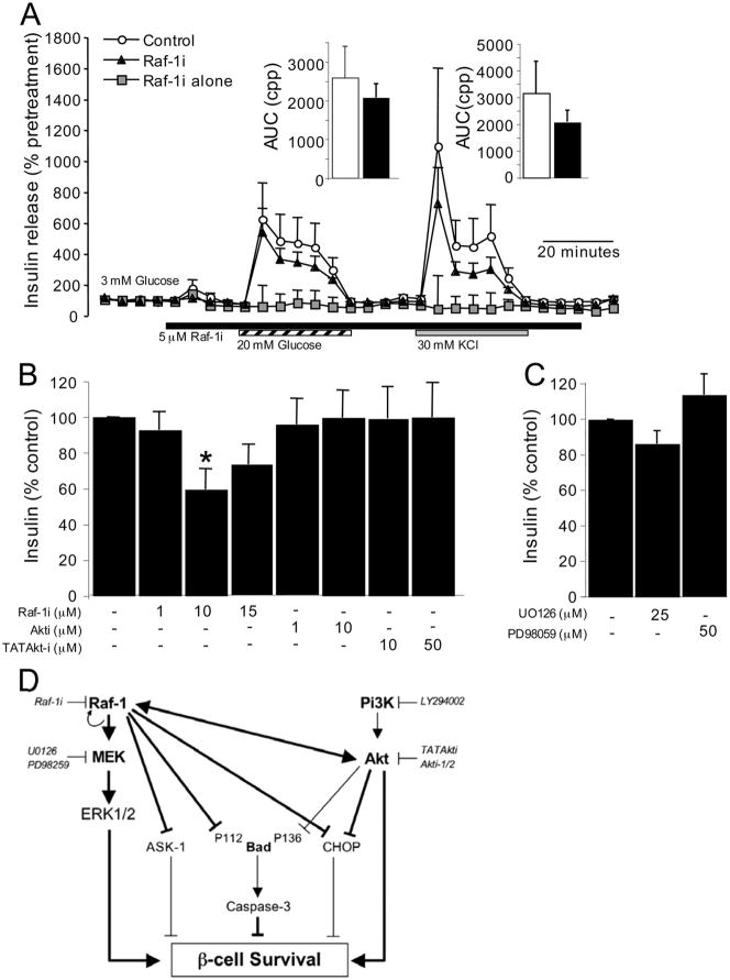
The serine threonine kinase Raf-1 plays a protective role in many cell types, but its function in pancreatic beta-cells has not been elucidated. In the present study, we examined whether primary beta-cells possess Raf-1 and tested the hypothesis that Raf-1 is critical for beta-cell survival. Using reverse transcriptase-PCR, Western blot, and immunofluorescence, we identified Raf-1 in human islets, mouse islets, and in the MIN6 beta-cell line. Blocking Raf-1 activity using a specific Raf-1 inhibitor or dominant-negative Raf-1 mutants led to a time- and dose-dependent increase in cell death, assessed by real-time imaging of propidium iodide incorporation, TUNEL, PCR-enhanced DNA laddering, and Caspase-3 cleavage. Although the rapid increase in apoptotic cell death was associated with decreased Erk phosphorylation, studies with two Mek inhibitors suggested that the classical Erk-dependent pathway could explain only part of the cell death observed after inhibition of Raf-1. An alternative Erk-independent pathway downstream of Raf-1 kinase involving the pro-apoptotic protein Bad has recently been characterized in other tissues. Inhibiting Raf-1 in beta-cells led to a striking loss of Bad phosphorylation at serine 112 and an increase in the protein levels of both Bad and Bax. Together, our data strongly suggest that Raf-1 signaling plays an important role regulating beta-cell survival, via both Erk-dependent and Bad-dependent mechanisms. Conversely, acutely inhibiting phosphatidylinositol 3-kinase Akt had more modest effects on beta-cell death. These studies identify Raf-1 as a critical anti-apoptotic kinase in pancreatic beta-cells and contribute to our understanding of survival signaling in this cell type.


Figures

FIGURE 1
FIGURE 1. Expression of Raf isoforms in pancreatic β-cells
A, total RNA was extracted from mouse islets and MIN6 cells and gene expression levels were analyzed by semi-quantitative reverse transcriptase-PCR (n = 3). B, Raf-1 protein expression was detected in human and mouse islets as well as in the MIN6 cell line using Western blot (n = 3). C, immunofluorescence imaging of Raf-1 in mouse β-cells, human β-cells, and MIN6 dispersed islet cells showing Raf-1 localization in the cytoplasm and the nucleus (>1000 cells examined). Scale bar, 5 μm. DAPI, 4′,6-diamidino-2-phenylindole.
FIGURE 2
FIGURE 2. Inhibition of endogenous Raf-1 signaling in primary mouse islet cells and MIN6 cells causes β-cell death
Rapid increase in propidium iodide (PI) incorporation in dispersed mouse islet cells (A) and MIN6 cells (B) treated with the specific inhibitor of Raf-1 kinase, Raf-1i (n = 9). Raf-1i increases β-cell death in a dose-and time-dependent manner. Raf-1i also caused β-cell apoptosis assessed by TUNEL staining of mouse islet β-cells (C) and MIN6 cells within 24 h (D). C, dispersed mouse β-cells stained for insulin are red, TUNEL positive cells are green, and cell nuclei stained with 4′,6-diamidino-2-phenylindole are blue. D, TUNEL-positive MIN6 cells are green and nuclei are blue. TUNEL staining experiments were repeated independently with similar results using dispersed human and mouse islets (≥8,168 cells examined for each condition) and MIN6 cells (≥16,061 cells for each condition). The ratio of TUNEL positive cells/total cells examined is shown as an inset. E, cleaved Caspase-3 protein levels in MIN6 cells treated with Raf-1i for 6 h (n = 3). F, quantification of cleaved Caspase-3 protein levels corrected to control. G, Raf-1i induces DNA laddering in intact mouse islets (n = 3). Asterisk denotes significant difference (p < 0.05) between the control and treatment.
FIGURE 3
FIGURE 3. Roles of Erk and Mek in Raf-1 inhibitor-induced β-cell death
A, phosphorylated and total Erk levels in MIN6 cells treated with Raf-1i for 6 h (n = 3). B, quantification of phosphorylated Erk to total Erk protein levels normalized to control. C, quantification of total Erk/β-Actin protein levels normalized to control. Moderate increase in propidium iodide incorporation in dispersed mouse islet cells (D) and MIN6 cells (E) treated with Mek inhibitors, UO126 and PD98059. F, phosphorylated and total Erk levels in MIN6 cells treated with UO126 and PD98059 for 3 h (n = 3). G, quantification of phosphorylated Erk to total Erk protein levels normalized to control. H, quantification total Erk/β-Actin ratio normalized to control. Asterisk denotes significant difference (p < 0.05) between the control and treatment.
FIGURE 4
FIGURE 4. Effects of Raf-1 inhibitor on Bad, Bcl-2, and Bax
A, phosphorylated Bad (Ser112) and total Bad in MIN6 cells treated with Raf-1i for 6 h (n = 3). B, quantification of the ratio of phosphorylated Bad Ser112 to total Bad protein (normalized to control). C, quantification of total Bad/β-Actin protein ratio normalized to control. D, Raf-1i increased Bax protein levels in MIN6 cells treated for 6 h (n = 3). E, quantification of total Bax/β-Actin protein ratio normalized to control. F, Bcl-2 protein levels/β-Actin protein normalized to the control (n = 3). G, quantification of total Bcl-2/β-Actin protein levels normalized to control. H, Bcl-2/Bax ratio. Asterisk denotes significant difference (p < 0.05) between the control and treatment.
FIGURE 5
FIGURE 5. Localization of Raf-1-Gfp fusion proteins in MIN6 cells
Widefield, deconvolution fluorescence imaging of Raf-1-Gfp fusion proteins and mitochondrial-targeted red fluorescent protein (DsRed-Mito) co-transfected into MIN6 cells. A, DsRed-Mito colocalizes strongly with Raf-1-Gfp, modestly with Raf-151–220-Gfp, but not with Raf-151–131-Gfp. B, Pearson correlation between Raf-1-Gfp fusion proteins and DsRed-Mito or Hoechst DNA dye (nucleus) were calculated as described under “Experimental Procedures.” Scale bar, 2 μm.
FIGURE 6
FIGURE 6. Expressed Raf-1-Gfp fusion proteins inhibit Raf-1-mediated Erk activation and induces MIN6 cells β-cell death
A, sample FACS scatter plots showing non-GFP expressing MIN6 cells and Gfp-positive cells. B, phosphorylated Erk and cleaved Caspase-3, total Erk, and β-Actin in FACS-enriched Gfp-positive MIN6 cells (n = 3). C, quantification of phosphorylated Erk/total Erk protein ratio normalized to control. D, quantification of cleaved Caspase-3 protein/β-Actin ratio normalized to control. E, expression of dominant-negative Raf-1 fusion proteins caused an increased in propidium iodide incorporation in MIN6 cells. Asterisk denotes significant difference (p < 0.05) between the control and treatment.
FIGURE 7
FIGURE 7. The relative roles of Raf-1 and PI 3-kinase/Akt signaling on ER stress and in β-cell apoptosis
Rapid increase in propidium iodide incorporation in dispersed mouse islet cells (A) and MIN6 cells (B) treated with specific inhibitors of Raf-1 kinase (Raf-1i), PI 3-kinase (LY294002), or Akt kinase (Akti-1/2 and TATAkt-in). Like Raf-1i, LY294002 and Akti-1/2 increased β-cell death in a dose- and time-dependent manner, suggesting that all three kinases play a role in β-cell survival (n = 6). C, effects of Raf-1i and Akti-1/2 on the expression of CHOP, an ER stress marker, and cleavage of caspase-3 in MIN6 cells treated for 3 h (n = 3). C, phosphorylated and total Erk and phosphorylated (Ser112) Bad and total Bad levels in MIN6 cells treated with Raf-1i and Akti-1/2 for 3 h (n = 3). Quantification of Chop/β-Actin ratio (D) and cleaved Caspase-3/β-Actin ratio (E) normalized to control. F, quantification of Bad Ser112/total Bad protein ratio normalized to the control. G, quantification of total Bad/β-Actin ratio corrected to control. H, quantification of phosphorylated and the total Erk level ratio normalized to control. Asterisk denotes a significant difference (p < 0.05) between the control and treatment.
FIGURE 8
FIGURE 8. Raf-1 inhibitor reduces Akt activity and Raf-1 protein levels
A, phosphorylated Akt (Ser473) and total Akt in MIN6 cells treated with Raf-1i for 3 h (n = 3). B, quantification of phosphorylated Akt (Ser473) and total Akt protein levels normalized to the control. C, quantification of total Akt normalized to the control. D, Raf-1i and Akti-1/2 both reduced total Raf-1 protein levels in MIN6 cells. E, quantification as shown (n = 3). Note that this is the same β-Actin loading control shown in Fig. 7C. F, additive effects of blocking both Raf-1 and Akt on β-cell death. Rapid increase in propidium iodide (PI) incorporation in the presence of Raf-1i alone, or the combination of Raf-1i and Akti-1/2 MIN6 cells (n = 3). Asterisk denotes a significant difference (p < 0.05) between the control and treatment.
FIGURE 9
FIGURE 9. Effects of Raf-1 inhibitor on insulin secretion in primary islets and MIN6 cells
A, groups of 100 mouse islets were perifused with Krebs-Ringer buffer containing 3 mM glucose. Islets were exposed to 20 mM glucose (striped bar), 30 mM KCl (gray bar), in the presence or absence of 5 μM Raf-1i (black bar). Control islets (open circles) were exposed to glucose and KCl without Raf-1i. Islets treated with Raf-1i (closed triangle) were also exposed to glucose and KCl. Some islets were exposed to Raf-1i alone (gray squares) without glucose or KCl. Values are normalized to the pretreatment levels of insulin secretion to compensate for uneven numbers of islets in each column (n = 4). Area under the curve (AUC) was measured as the cumulative percent pre-treatment (cpp) and shown as insets. Insulin levels were measured in conditioned media of MIN6 cells treated with Raf-1i, Akti-1/2, UO126, and PD98059 for 3 h (n = 3) (B and C). Asterisk denotes a significant difference (p < 0.05) between the control and treatment. D, simplified diagram of the pro-survival Raf-1/Erk and PI 3-kinase/Akt signaling network in the β-cell. Major pro-survival factors in the β-cell activate a series of common signaling events that can be broadly divided into two arms, namely the PI 3-kinase/Akt and the Raf-1/Erk pathways. Activation of Raf-1 can lead to the induction of Erk activity or reduction of the pro-apoptotic effects of Ask-1, Bad, and Chop. Akt signaling prevents β-cell apoptosis in part through Foxo1, a negative regulator of β-cell survival (not shown) and by regulating other pro-apoptotic proteins (Bad and Chop). Raf-1 and Akt cross-talk at multiple levels and their signals are interdependent and coordinated to maintain β-cell survival. Interactions examined in the present study are illustrated by the thick lines, whereas interactions addressed by previous research are indicated by thin lines.

References

    1. Augstein P, Stephens LA, Allison J, Elefanty AG, Ekberg M, Kay TW, Harrison LC. Mol Med. 1998;4:495–501. - PMC - PubMed
    1. Donath MY, Halban P. Diabetologia. 2004;47:581–589. - PubMed
    1. Ramachandran S, Desai NM, Goers TA, Benshoff N, Olack B, Shenoy S, Jendrisak MD, Chapman WC, Mohanakumar T. Am J Transplant. 2006;6:1696–1703. - PubMed
    1. Kulkarni RN, Bruning JC, Winnay JN, Postic C, Magnuson MA, Kahn CR. Cell. 1999;96:329–339. - PubMed
    1. Hashimoto N, Kido Y, Uchida T, Asahara S, Shigeyama Y, Matsuda T, Takeda A, Tsuchihashi D, Nishizawa A, Ogawa W, Fujimoto Y, Okamura H, Arden KC, Herrera PL, Noda T, Kasuga M. Nat Genet. 2006;38:589–593. - PubMed

Publication types

MeSH terms

LinkOut - more resources