. 2008 Mar;22(3):760-71.
doi: 10.1210/me.2007-0330. Epub 2007 Nov 15.

Adiponectin activates adenosine monophosphate-activated protein kinase and decreases luteinizing hormone secretion in LbetaT2 gonadotropes

Affiliations

Adiponectin activates adenosine monophosphate-activated protein kinase and decreases luteinizing hormone secretion in LbetaT2 gonadotropes

Min Lu et al. Mol Endocrinol. 2008 Mar.

Abstract

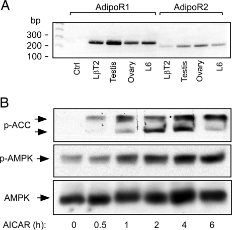
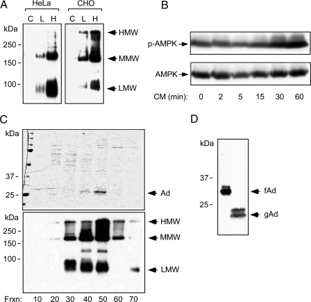
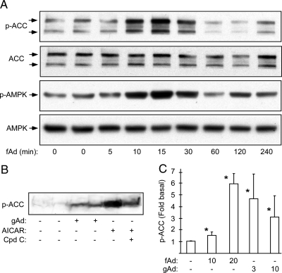
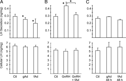
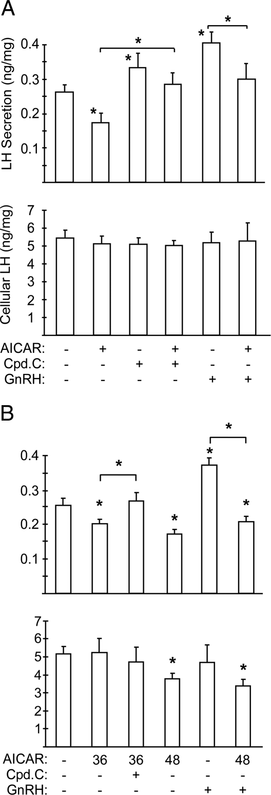
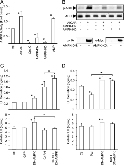
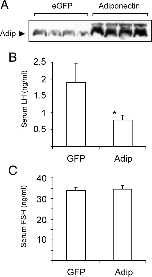
Metabolic dysregulation is associated with reproductive disorders, but the underlying mechanisms are not clearly understood. Adiponectin is an adipocyte-derived secretory factor that improves insulin sensitivity. Results from animal models indicate that overexpression of adiponectin impairs female fertility. We hypothesized that adiponectin regulates reproduction by altering the hypothalamic-pituitary axis. Mouse LbetaT2 immortalized gonadotrope cells express both adiponectin receptors 1 and 2. Adiponectin increases phosphorylation of AMP-activated protein kinase (AMPK), a downstream target of adiponectin receptors, and reduces basal and GnRH-stimulated LH secretion, acutely. The repression of LH secretion can be mimicked by 5-aminoimidazole-4-carboxamide-1-beta-riboside, an AMP analog, suggesting the involvement of AMPK. A dominant-negative AMPK mutant or compound C, a selective AMPK inhibitor, potentiates basal LH secretion and abolishes the inhibitory effect of adiponectin. Chronic activation of AMPK by 5-aminoimidazole-4-carboxamide-1-beta-riboside decreases cellular LH levels, and expression of dominant-negative AMPK increases cellular LH levels, suggesting a second effect of AMPK to regulate LH synthesis. Lastly, intravenous injection of an adenovirus expressing adiponectin into male mice reduces serum LH levels without changing FSH levels. In conclusion, our results suggest that adiponectin decreases LH secretion in pituitary gonadotropes in an AMPK-dependent manner.


Figures

Figure 1
Figure 1
Detection of AdipoR1 and -2 and Downstream Signaling Pathways in LβT2 Gonadotropes A, LβT2 cells express AdipoR1 and R2. Total RNA extracted from LβT2 gonadotropes, L6 rat skeletal muscle cells, mouse testis, and ovary were subjected to RT-PCR for mouse and rat AdipoR1 (270 bp) or -R2 (220 bp). B, AICAR stimulates AMPK and ACC phosphorylation. LβT2 cells were treated with 1 mm AICAR for the times indicated. Total cell lysates were extracted and subjected to immunoblotting. Phospho-AMPKα (Thr172), phospho-ACC (Ser79), and total AMPKα were detected by immunoblotting. Ctrl, Control.
Figure 2
Figure 2
Purification of Recombinant Adiponectin from Mammalian Cells A, Adiponectin expression in HeLa or CHO cells. Cells infected with an adenovirus- expressing mouse adiponectin were used to produce recombinant full-length adiponectin (C, L, and H indicate control, low dose, and high virus dose, respectively). Conditioned media (48 h) were subjected to nondenaturing, nonreducing SDS-PAGE. Multimerization of adiponectin was detected by immunoblotting. Arrows indicate the high-, medium (hexamer)-, and low-molecular weight (trimer) multimeric forms. B, Infected cells secrete adiponectin. LβT2 cells were serum starved and treated with conditioned media (CM) for the times indicated. Phosphorylation of AMPK and total AMPK α-subunit were detected by immunoblotting as before. C, Purifcation of recombinant full-length adiponectin from adenovirally infected CHO cells. Recombinant proteins were separated by fast protein liquid chromatography. Protein samples from different fractions, as indicated, were subjected to SDS-PAGE and Coommassie blue staining to determine the purity (top panel). The same samples were subjected to a nonreducing and nondenaturing gel followed by immunoblotting to determine the higher-order structure of adiponectin (bottom panel). Arrows indicate the high (HMW), the medium (MMW), and the low molecular (LMW) form of the recombinant adiponectin. D, Recombinant mouse full-length adiponectin (fAd) and human globular adiponectin (gAd) (a kind gift from Xencor, Inc.) were subjected to SDS-PAGE and detected by immunoblotting.
Figure 3
Figure 3
Purified Recombinant Adiponectin Activates AMPK in LβT2 Cells A, Full-length adiponectin stimulates AMPK. LβT2 cells were serum starved overnight and then treated with 20 μg/ml recombinant mouse full-length adiponectin (fAd) for the times indicated. Phospho-AMPKα, phospho-ACC, and total AMPKα were detected by immunoblotting. B, Globular adiponectin stimulates ACC phosphorylation. LβT2 cells were serum starved overnight followed by treatments with 3 μg/ml of human globular adiponectin (gAd) for 5 min. Treatment with 1 mm AICAR for 1 h served as a positive control. For inhibition of AICAR-induced AMPK activation, compound C (20 μm) was added for 30 min before AICAR treatment. C, Dose-dependent phosphorylation of ACC by fAd (10 and 20 μg/ml) and gAd (3 and 10 μg/ml). Data shown are the fold induction of ACC phosphorylation (mean ± sd) from three experiments. Asterisk indicates statistical significance (P < 0.05). Cpd C, Compound C.
Figure 4
Figure 4
Secretion of LH from LβT2 Cells Is Reduced by Adiponectin A, Adiponectin acutely inhibits LH secretion. LβT2 cells were serum starved, washed, and then treated with vehicle control (Ctl), 20 μg/ml full-length adiponectin (fAd,), or 10 μg/ml globular adiponectin (gAd) for 30 min. Secreted LH and cellular LH were assayed in conditioned media or cell lysates. LH levels were normalized to total cellular protein concentrations, and LH secretion (ng) per mg of cell lysate is shown. Experiments were performed in triplicate. Data are shown as mean ± sd from three experiments. B, Adiponectin inhibits GnRH-stimulated secretion. LβT2 cells were serum starved, washed, and then treated with 20 μg/ml full-length adiponectin in the presence or absence of 100 nm GnRH for 30 min. Secreted LH and cellular LH were assayed in conditioned media and cell lysates as above. C, Chronic adiponectin treatment has no effect on LH secretion. LβT2 cells were cultured and serum starved in the presence of 10 μg/ml globular adiponectin or 20 μg/ml full-length adiponectin for 48 h. Secreted LH and cellular LH were assayed as above. Asterisks indicate statistical significance (P < 0.05) vs. control or between conditions connected by bars.
Figure 5
Figure 5
AMPK Stimulation Decreases LH Secretion in LβT2 Cells A, Artificial activation of AMPK inhibits LH secretion. LβT2 cells were serum starved and then treated with 1 mm AICAR for 2 h with or without 20 μm compound C (Cpd.C) or 100 nm GnRH for 30 min. LH levels were normalized to cellular protein concentrations, and LH secretion (ng) per mg of cell lysates is shown. B, Chronic activation of AMPK inhibits LH secretion and synthesis. LβT2 cells were cultured and serum starved in the presence of 200 μm AICAR for 36 or 48 h. Compound C (20 μm) was added 1 h before the secretion assay and 100 nm GnRH was added for the final 30 min. Secreted LH and cellular LH were assayed as above. Experiments were performed in triplicate. Data are shown as mean ± sd from three experiments. Asterisks indicate statistical significance (P < 0.05) vs. control or between conditions connected by bars.
Figure 6
Figure 6
AMPK Mediates the Inhibitory Effect of Adiponectin on LH Secretion A, In vitro kinase activity of mutant AMPKs. LβT2 cells were infected with adenoviruses expressing either a dominant-negative form of AMPKα1 (AMPK-DN) or a kinase-dead form of AMPKα2 (AMPK-KD) for 48 h. Cells were also treated with 20 μm compound C (Cpd.C) or 1 mm AICAR for 4 h. AMPK activity was determined by kinase assay using SAMS peptide. A kinase reaction in the presence of 300 μm AMP serves as a positive control. B, Mutant AMPKs block endogenous AMPK activity in cells. CHO cells were infected with AMPK-DN or AMPK-KD. Cells were treated with 1 mm AICAR for 2 h, and phospho-ACC was detected by immunoblotting whole-cell extracts. Extracts of uninfected and infected cells were immunoblotted for the HA or c-Myc tags to demonstrate mutant protein expression (lower panels). C, A dominant-negative AMPK mutant increases LH secretion and synthesis. LβT2 cells were infected with dominant-negative AMPK adenovirus (AMPK-DN) or control virus (GFP). Cells were serum starved 32 h later for an additional 16 h. Cells were then washed and subjected to secretion in a 30-min period with or without 100 nm GnRH. Secreted LH and cellular LH were determined, and LH levels (ng) per mg of cell lysates are shown. D, A dominant-negative AMPK mutant blocks the acute effect of adiponectin. Infected cells were treated with 20 μg/ml adiponectin (fAd) for 30 min. Secreted and cellular LH levels were determined. Experiments were performed in triplicate. Data are shown as mean ± sd from three experiments. Asterisks indicate statistical significance (P < 0.05) vs. control or between conditions connected by bars. HA, Hemagglutinin; Ctl, control.
Figure 7
Figure 7
Adiponectin Regulates Serum LH In Vivo A, Adiponectin expression in infected mice. Male C57BL/6J mice (8 wk old) were injected iv with either GFP adenovirus (eGFP) (n = 16) or adiponectin adenovirus (adiponectin) (n = 17). All mice were killed 7 d later, and serum samples were subjected to immunoblotting to determine adiponectin levels. B, Adiponectin expression reduces serum LH. C, Adiponectin expression has no effect on serum FSH. Graphs show serum gonadotropin levels (mean ± sem). Asterisk indicates statistical significance (P < 0.05) vs. GFP-infected mice. Adip, Adiponectin.

References

    1. Schneider JE 2004 Energy balance and reproduction. Physiol Behav 81:289–317 - PubMed
    1. Wade GN, Jones JE 2004 Neuroendocrinology of nutritional infertility. Am J Physiol 287:R1277–R1296 - PubMed
    1. Mitchell M, Armstrong DT, Robker RL, Norman RJ 2005 Adipokines: implications for female fertility and obesity. Reproduction (Cambridge, England) 130:583–597 - PubMed
    1. Trujillo ME, Scherer PE 2006 Adipose tissue-derived factors: impact on health and disease. Endocr Rev 27:762–778 - PubMed
    1. Zhang Y, Proenca R, Maffei M, Barone M, Leopold L, Friedman JM 1994 Positional cloning of the mouse obese gene and its human homologue. Nature 372:425–432 - PubMed

Publication types

MeSH terms