. 2007 Nov 27;104(48):19011-6.
doi: 10.1073/pnas.0706696104. Epub 2007 Nov 15.

Protein phosphatase 2A regulates life and death decisions via Akt in a context-dependent manner

Affiliations

Protein phosphatase 2A regulates life and death decisions via Akt in a context-dependent manner

Shaida Andrabi et al. Proc Natl Acad Sci U S A. .

Abstract

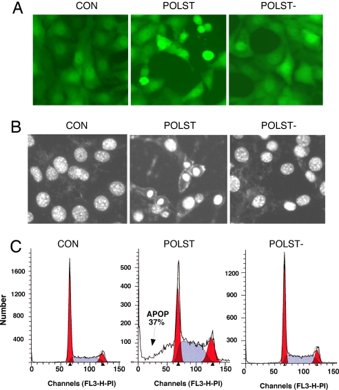
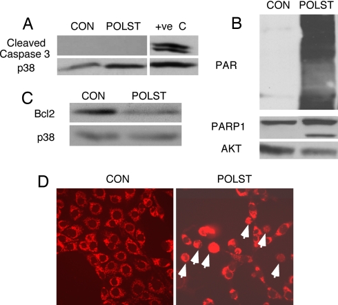
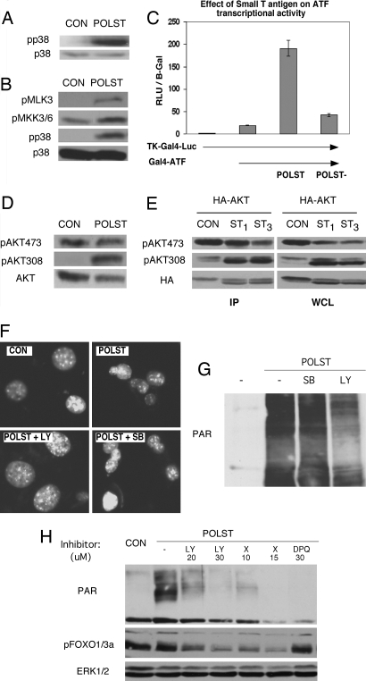
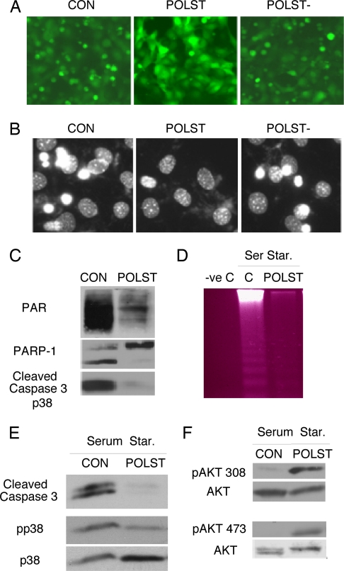
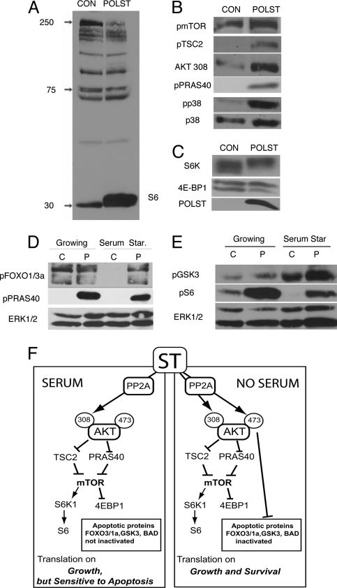
Here, we show how targeting protein phosphatase 2A (PP2A), a key regulator of cellular protein phosphorylation, can either induce or prevent apoptosis depending on what other signals the cell is receiving. The oncoprotein polyoma small T interacts with PP2A to regulate survival. In the presence of growth factors, small T induces apoptosis. Akt activity, which usually promotes survival, is required for this death response, because inhibitors of Akt or PI3 kinase protect cells from death. The activation of Akt under these conditions is partial, characterized by T308 phosphorylation but not S473 phosphorylation. In the absence of growth factors, small T protects from cell death. Here, small T uses PP2A to promote phosphorylation of Akt on both T308 and S473. This effect results in a different pattern of phosphorylation of Akt substrates and shifts Akt from a proapoptotic (presence of growth factors) to an antiapoptotic mode (absence of growth factors). An intriguing possibility is that Akt phosphorylation could be therapeutically disregulated to decrease the survival of cancer cells.


Conflict of interest statement

The authors declare no conflict of interest.

Figures

Fig. 1.
Fig. 1.
Polyoma small T is proapoptotic in presence of serum: NIH 3T3 cells were infected with retrovirus expressing GFP only (CON), expressing GFP and wild-type small T (POLST), or GFP and mutant small T defective in PP2A binding (POLST−). (A) Morphology of cells growing in 10% calf serum seen by fluorescence microscopy (×20 magnification). (B) Nuclear morphology seen by Hoechst 33342 staining and fluorescence microscopy (×20 magnification). (C) FACS analysis of cells growing in 10% calf serum stained with propidium iodide. Cells with subG1 DNA content (37% of the population) are indicated by the arrow.
Fig. 2.
Fig. 2.
Characterization of apoptosis induced by small T. (A) Blotting of cleaved caspase 3 in control or small T-expressing cells growing in 10% calf serum. Extract from NIH 3T3 cells serum-starved overnight was used as a positive control (+ve C). Total p38 was used as a loading control. (B) Blotting of cell extracts with antibody against Poly ADP Ribose (PAR) or PARP-1. Total Akt was used as a loading control. (C) Blotting of cell extracts with antibody against Bcl2. Total p38 was used as loading control. (D) Translocation of apoptosis inducing factor (AIF) from mitochondria to the nucleus in polyoma small T-expressing, but not control cells, detected by immunofluorescence stained with Trit-C by using antibody against AIF.
Fig. 3.
Fig. 3.
Small T regulation of cell signaling in growing conditions. NIH 3T3 cells infected with retrovirus expressing GFP only (CON) or expressing GFP and wild-type small T (POLST) were grown in 10% calf serum. (A) Cell extracts were blotted with antibody specific for phospho p38 (pp38) or total p38 (p38). (B) Cell extracts of control or small T-expressing cells were blotted with anti-phospho MLK3 or MKK3/6. Phospho p38 and p38 blots are shown as controls. (C) Effect of small T on the activity of ATF2, a downstream target of p38. ATF2 activity was measured in a reporter assay by cotransfection of TKGal4 luciferase, GAL4-ATF2 with wild-type small T (POLST), PP2A− small T (POLST−), or empty vector as shown. (D) Cell extracts were blotted with antibody specific for phosphorylation of T308 or phosphorylation of S473 to assess activation of Akt. Anti-Akt was used to determine the amount of protein. (E) Cells (293T) were transfected with HA-Akt and control (CON), 1 μg (ST1), or 3 μg (ST3) of small T plasmid. Extracts (Right) or HA-Akt immunoprecipitates (Left) were made 48 h after transfection. Blotting with antibody specific for phosphorylation of T308 or phosphorylation of S473 on Akt was used to assess activation, and total transfected HA-Akt was determined by using HA11. (F) Nuclear morphology seen by Hoechst 33342 staining and fluorescence microscopy (×20 magnification) in untreated cells or in cells treated with PI3 kinase inhibitor LY294002 (24 h, 20 μM) or with p38 inhibitor SB203580 (24 h, 20 μM). (G) Small T-expressing cells growing in 10% serum were treated with PI3 kinase inhibitor LY294002 (20–24 h, 20 μM) or p38 inhibitor SB203580 (20–24 h, 20 μM). Extracts from control and treated cells were blotted with PAR antibody. (H) Small T-expressing cells growing in 10% serum were treated with specific Akt inhibitor AktX (10 μM and 15 μM), PI3 kinase inhibitor LY294002 (20 μM and 30 μM), or PARP-1 inhibitor DPQ (30 μM) for 16–20 h. Extracts from control and treated cells were blotted with PAR antibody. The inhibition of Akt activity was indicated by blotting with phosphospecific FOXO1/3a antibody, and ERK1/2 served as a loading control.
Fig. 4.
Fig. 4.
Polyoma small T is prosurvival under serum-starvation conditions. (A) Morphology of control, wild type or PP2A− mutant small T-expressing cells growing in the absence of serum overnight (16–24 h) as seen by fluorescence microscopy for GFP (×10 magnification). (B) Nuclear morphology seen by Hoechst 33342 staining and fluorescence microscopy (×20 magnification) of cells treated as in A. (C) Control and small T-expressing cells were serum starved overnight. Cells were protected from death by small T as shown by blotting of cell extracts for PAR, PARP-1, and caspase 3 activation. (D) Polyoma small T suppressed DNA laddering caused by serum starvation. DNA was extracted from cells growing in 10% serum (-ve C) or starved overnight (control or small T). Equal amounts of DNA were loaded on a 2% gel and stained with ethidium bromide. (E) Cell extracts from control or small T-expressing cells starved overnight were blotted with antibody specific for caspase 3, phospho p38 (pp38), or total p38 (p38). (F) Cell extracts from control or small T-expressing cells starved overnight were blotted with anti-p308 Akt antibody (Upper) and anti-pS473 Akt antibody (Lower). Anti-Akt was used to determine the amount of Akt protein.
Fig. 5.
Fig. 5.
Effects of small T on phosphorylation of Akt pathway substrates. (A) Phosphorylation of Akt substrates was assessed by blotting of extracts from control or small T-expressing cells grown in 10% serum. Akt substrate antibody that recognizes the phosphorylated consensus Akt substrate site (RXRXXS/T) was used for blotting. The identification of one band as S6 was confirmed with phosphospecific S6 antibody (data not shown). (B) Phosphorylation of mTOR pathway proteins mTOR, TSC2, and PRAS40 was determined by using phosphospecific antibody for each substrate. Phosphorylation of Akt 308 and p38 was used as a control for small T-induced signaling, and total p38 was used as a loading control. Cells were grown in 10% serum. (C) Small T expression enhances S6K and 4EBP1 phosphorylation as demonstrated by reduced gel mobilities shown in blots with anti S6K and 4EBP1 antibodies of small T-expressing compared with control cells. Cells were grown in 10% serum. (D) Some differences in signaling that change from growing conditions (in presence of serum) and serum starvation. Extracts were made from cells growing in 10% serum or after serum starvation (16–24 h). Extracts from control (C) or polyoma (P) small T-expressing cells were blotted for phosphorylation of FOXO1/3a and PRAS40 by using antibodies that detect Akt phosphorylation sites on these proteins. Total ERK1/2 was used as loading control. (E) Extracts from control (C) or polyoma (P) small T-expressing cells as in D were blotted for phospho GSK3α/β and phospho S6 by using phosphospecific antibodies. ERK1/2 was used as loading control. (F) Proposed model showing the signaling of small T in growing and serum-starvation conditions. Branch 1 shows that in growing conditions (presence of serum), small T shifts the Akt signaling to translational pathway only, whereas proteins involved in apoptosis like FOXO1/3a, GSK3, and BAD are not inactivated. In serum starvation (Branch 2), small T causes Akt phosphorylation at both S473 and T308, which activates Akt fully and causes phosphorylation of relevant substrates including FOXO1/3a, GSK3, and BAD, thereby giving protection against apoptotic signals.

References

    1. Engelman JA, Luo J, Cantley LC. Nat Rev Genet. 2006;7:606–619. - PubMed
    1. Datta SR, Brunet A, Greenberg ME. Genes Dev. 1999;13:2905–2927. - PubMed
    1. Maurer U, Charvet C, Wagman AS, Dejardin E, Green DR. Mol Cell. 2006;21:749–760. - PubMed
    1. Gao T, Furnari F, Newton AC. Mol Cell. 2005;18:13–24. - PubMed
    1. Brognard J, Sierecki E, Gao T, Newton AC. Mol Cell. 2007;25:917–931. - PubMed

Publication types

MeSH terms

LinkOut - more resources