Comparative Study
. 2008 Feb;324(2):463-74.
doi: 10.1124/jpet.107.132332. Epub 2007 Nov 15.

Molecular basis for agonist selectivity and activation of the orphan bombesin receptor subtype 3 receptor

Affiliations
Comparative Study

Molecular basis for agonist selectivity and activation of the orphan bombesin receptor subtype 3 receptor

Nieves Gonzalez et al. J Pharmacol Exp Ther. 2008 Feb.

Abstract

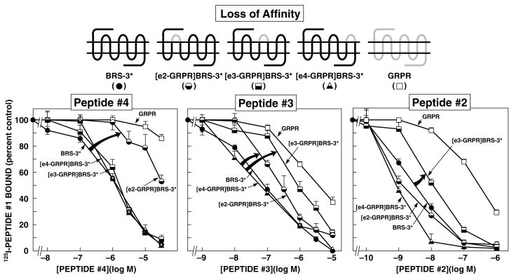
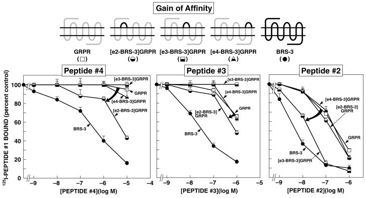
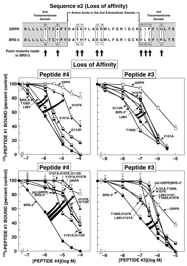
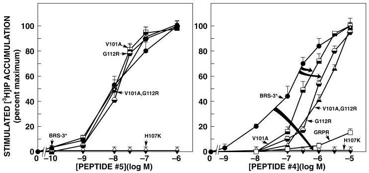
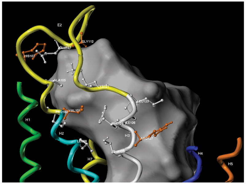
Bombesin receptor subtype (BRS)-3, a G-protein-coupled orphan receptor, shares 51% identity with the mammalian bombesin (Bn) receptor for gastrin-releasing peptide. There is increasing interest in BRS-3 because it is important in energy metabolism, glucose control, motility, and tumor growth. BRS-3 has low affinity for all Bn-related peptides; however, recently synthetic high-affinity agonists, [d-Tyr(6)/d-Phe(6),betaAla(11),Phe(13),Nle(14)]Bn-(6-14), were described, but they are nonselective for BRS-3 over other Bn receptors. Based on these peptides, three BRS-3-selective ligands were developed: peptide 2, [d-Tyr(6)(R)-3-amino-propionic acid(11),Phe(13),Nle(14)]Bn(6-14); peptide 3, [d-Tyr(6),(R)-Apa(11),4Cl-Phe(13),Nle(14)]Bn(6-14); and peptide 4, acetyl-Phe-Trp-Ala-His-(tBzl)-piperidine-3 carboxylic acid-Gly-Arg-NH(2). Their molecular determinants of selectivity/high affinity for BRS-3 are unknown. To address this, we used a chimeric/site mutagenesis approach. Substitution of extracellular domain 2 (EC2) of BRS-3 by the comparable gastrin-releasing peptide receptor (GRPR) domain decreased 26-, 4-, and 0-fold affinity for peptides 4, 3, and 2. Substitution of EC3 decreased affinity 4-, 11-, and 0-fold affinity for peptides 2 to 4. Ten-point mutations in the EC2 and adjacent transmembrane regions (TM2) 2 and 3 of BRS-3 were made. His107 (EC2-BRS-3) for lysine (H107K) (EC2-GRPR) decreased affinity (25- and 0-fold) for peptides 4 and 1; however, it could not be activated by either peptide. Its combination with Val101 (TM2), Gly112 (EC2), and Arg127 (TM3) resulted in complete loss-of-affinity of peptide 4. Receptor-modeling showed that each of these residues face inward and are within 4 A of the binding pocket. These results demonstrate that Val101, His107, Gly112, and Arg127 in the EC2/adjacent upper TMs of BRS-3 are critical for the high BRS3 selectivity of peptide 4. His107 in EC2 is essential for BRS-3 activation, suggesting amino-aromatic ligand/receptor interactions with peptide 4 are critical for both binding and activation. Furthermore, these result demonstrate that even though these three BRS-3-selective agonists were developed from the same template peptide, [d-Phe(6),betaAla(11),Phe(13),Nle(14)]Bn-(6-14), their molecular determinants of selectivity/high affinity varied considerably.


Figures

Figure 1
Figure 1
Affinities of peptides#2–4 for BRS-3*, extracellular chimeric BRS-3*s, and wild-type GRPR expressed in CHOP cells (loss-of-affinity chimeras). The diagrams of the chimeric receptors formed are shown at the top. The chimeric BRS-3*s ([R127Q]BRS-3) were formed by replacing each of the extracellular domains of BRS-3 one at a time by the comparable GRPR extracellular domain. Results are expressed as the percentage of the saturable binding seen with no peptide#2-4 present that occurred when the indicated concentration of peptide#2-4 was present. Each point is the mean from at least 5 separate experiments, and in each experiment each point was measured in duplicate. EC2-, e3-, and e4-GRPR refer to the substitution of this extracellular domain of the GRPR for the comparable extracellular domain in BRS-3*. The arrows indicate large changes in affinity from BRS-3*.
Figure 2
Figure 2
Affinities of peptides#2-4 for wild-type GRPR, extracellular chimeric GRPRs, and wild-type BRS-3 expressed in CHOP cells (gain-of-affinity chimeras). Diagrams of the chimeric receptors formed are shown at the top. The chimeric GRPRs were formed by replacing each of the extracellular domains of GRPR by the comparable domain of BRS-3 one at a time. Results are expressed as the percentage of the saturable binding seen with no peptide#2-4 present that occurred when the indicated concentration of peptide#2–4 was present. Each point is the mean from at least 5 separate experiments, and in each experiment each point was measured in duplicate. EC2-, e3-, and e4-BRS-3 refer to the substitution of this extracellular domain of BRS-3 for the comparable extracellular domain in GRPR. The arrows indicate large changes in affinity from the wild-type GRPR.
Figure 3
Figure 3
Effect of various point mutations alone or in combination in the 2nd extracellular domain and adjacent transmembrane domains BRS-3* on affinity for peptides#3 and #4. (Top panel) Alignment of amino acids sequences in the second extracellular domain or adjacent transmembrane regions of GRPR and BRS-3. The boxes indicate divergent amino acids between these two receptors in these regions. Shown are the ten BRS-3* mutants made to explore the importance of ten amino acid differences for determining peptide#4 and peptide#3 selectivity. Arrows indicate that position of the point mutations made in BRS-3* by substituting into BRS-3* the divergent amino acid from the comparable position in GRPR. (Middle and Bottom panel) Affinities of peptide#4 and peptide#3 for BRS-3*, second extracellular domain or adjacent TM or point and combination point mutants of BRS-3* expressed in CHOP cells (BRS-3* loss-of-affinity point and combination point mutants). Results are expressed as the percentage of the saturable binding seen with no peptide#3 or 4 present that occurred when the indicated concentration of peptide#3 or 4 was present. Each point is the mean from at least 5 separate experiments, and in each experiment each point was measured in duplicate. The arrows indicate large changes in affinity of the BRS-3* by the mutations. Abbreviations: BRS-3*-[R127Q]BRS-3; [EC2-GRPR]BRS-3 refers to the substitution of the 2nd extracellular domain from GRPR into the comparable location in BRS-3.
Figure 4
Figure 4
Ability of peptides#4 and #5 to stimulate increases in [3H]IP formation in second extracellular domain or adjacent transmembrane regions point and combination point mutants of GRPR, and BRS-3*-R-transfected Balb-3T3 cells. The different nature and mutant receptors (5.0x10⁴ cells/well) were loaded with myo-[2-3H]inositol as described in METHOS, washed, and incubated with the indicated concentrations of peptide#4 or #5 for 45 min at 37oC. Values are expressed as the percent of the total [3H]IP release stimulated by 1 μM peptide#5. Results are the means ±SEM from at least 5 experiments, and each point was determined in duplicate. The control and maximal value for BRS-3* were 1358±199 dpm and 3958±889 dpm (n=6).
Figure 5
Figure 5
Three dimensions model of the putative binding pocket involving the EC2 region of the hBRS-3-receptor. This model was based on the studies of bovine rhodopsin as described in METHODS. The interior of the extracellular solvent-accessible region of the receptor is indicated as a white shape. This central portion of the transmembrane receptor may accommodate and bind ligands. Solvent-accessible amino acid residues within interaction distance (4Å) of this binding pocket are shown as ball and stick models, with the residues identified in this work as important for binding colored orange [Val101, His107, Gly112 and Arg127]. Residues L98, V101, H107, E111, G112, which differed between BRS-3 and GRPR (Fig. 3) are within 4Å of the binding pocket.

References

    1. Akeson M, Sainz E, Mantey SA, Jensen RT, Battey JF. Identification of four amino acids in the gastrin-releasing peptide C receptor that are required for high affinity agonist binding. J Biol Chem. 1997;272:17405–17409. - PubMed
    1. Benya RV, Akeson M, Mrozinski J, Jensen RT, Battey JF. Internalization of the gastrin-releasing peptide receptor is mediated by phospholipase C-dependent and -independent processes. Mol Pharmacol. 1994;46:495–501. - PubMed
    1. Boyle RG, Humphries J, Mitchell T, Showell GA, Apaya R, Iijima H, Shimada H, Arai T, Ueno H, Usui Y, Sakaki T, Wada E, Wada K. The design of a new potent and selective ligand for the orphan bombesin receptor subtype 3 (BRS3) J Pept Sci. 2005;11:136–141. - PubMed
    1. Case DA, Cheatham TE, III, Darden T, Gohlke H, Luo R, Merz KM, Jr, Onufriev A, Simmerling C, Wang B, Woods RJ. The Amber biomolecular simulation programs. J Comput Chem. 2005;26:1668–1688. - PMC - PubMed
    1. Couvineau A, Rouyer-Fessard C, Maoret JJ, Gaudin P, Nicole P, Laburthe M. Vasoactive intestinal peptide (VIP)1 receptor. Three nonadjacent amino acids are responsible for species selectivity with respect to recognition of peptide histidine isoleucinemide. J Biol Chem. 1996;271:12795–12800. - PubMed

Publication types

LinkOut - more resources