. 2008 Apr;128(4):939-48.
doi: 10.1038/sj.jid.5701132. Epub 2007 Nov 15.

Isolation of pathogenic monoclonal anti-desmoglein 1 human antibodies by phage display of pemphigus foliaceus autoantibodies

Affiliations

Isolation of pathogenic monoclonal anti-desmoglein 1 human antibodies by phage display of pemphigus foliaceus autoantibodies

Ken Ishii et al. J Invest Dermatol. 2008 Apr.

Abstract

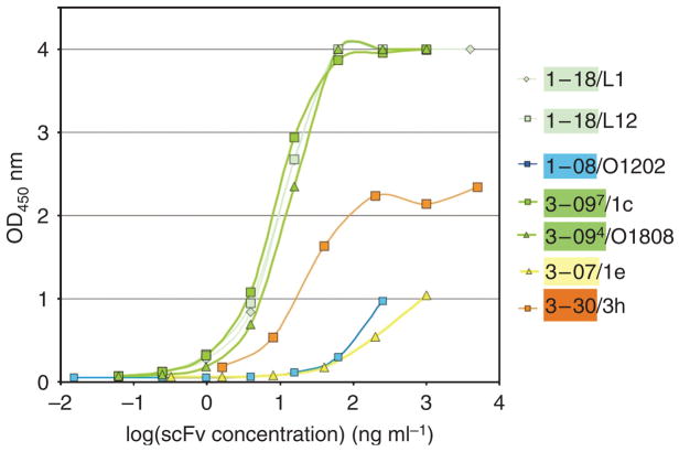
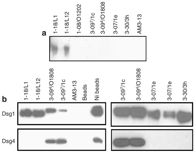
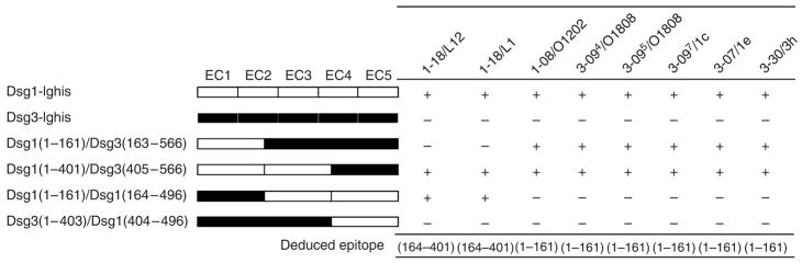
Pemphigus foliaceus (PF) is a blistering disease caused by autoantibodies to desmoglein 1 (Dsg1) that cause loss of epidermal cell adhesion. To better understand PF pathophysiology, we used phage display to isolate anti-Dsg1 mAbs as single-chain variable fragments (scFvs) from a PF patient. Initial panning of the library isolated only non-pathogenic scFvs. We then used these scFvs to block non-pathogenic epitopes and were able to isolate two unique scFvs, each of which caused typical PF blisters in mice or human epidermis models, showing that a single mAb can disrupt Dsg1 function to cause disease. Both pathogenic scFvs bound conformational epitopes in the N terminus of Dsg1. Other PF sera showed a major antibody response against the same or nearby epitopes defined by these pathogenic scFvs. Finally, we showed restriction of the heavy-chain gene usage of all anti-Dsg1 clones to only five genes, which determined their immunological properties despite promiscuous light-chain gene usage. These mAbs will be useful for studying Dsg1 function and mechanisms of blister formation in PF and for developing targeted therapies and tools to monitor disease activity.


Conflict of interest statement

CONFLICT OF INTEREST

Drs Ishii, Siegel, and Stanley have filed for a provisional patent on the antibodies described herein.

Figures

Figure 1
Figure 1. Binding curves of representative scFv antibodies against human Dsg1 by ELISA
Serial dilutions of representative scFvs were measured by Dsg1 ELISA. Clones 1-18/L1 and 1-18/L12 (light green lines) and 3-094/O18O8 and 3-097/1c (dark green lines) show similar binding curves. In contrast, clones 1-08/O12O2 and 3-07/1e showed weak binding to Dsg1 ELISA. Clone 3-30/3h exhibited intermediate binding capacity.
Figure 2
Figure 2. Indirect immunofluorescence of anti-Dsg1 scFvs on human skin
Clone 3-30/3h scFv antibodies stained the cell surface of keratinocytes throughout the human epidermis. Pretreatment of human skin with EDTA prevented cell surface staining by 3-30/3h. Clone 3-094/O18O8 showed cytoplasmic staining in the superficial layers of the epidermis.
Figure 3
Figure 3. Binding of anti-Dsg1 scFvs to denatured Dsg1 and to Dsg4
(a) The purified ectodomain of human Dsg1 with an E-tag (Dsg1-EHis), produced by baculovirus, was used as an immunoblot substrate. ScFv heavy-chain variable regions encoded by the gene VH1-18, but not other scFvs, bound denatured Dsg1 on immunoblots. (b) Recombinant human Dsg1 or Dsg4 containing an E-tag (Dsg1-EHis or Dsg4-EHis), produced by baculovirus, was immunoprecipitated with representative anti-Dsg1 scFvs, control scFv (AM3-13), or Talon metal affinity resin (Ni-beads) and detected on an immunoblot by anti-E tag antibody.
Figure 4
Figure 4. Epitope mapping with competition ELISA
Wild-type and domain-swapped extracellular domains of human Dsg1 and Dsg3 were produced by baculovirus and used as competitors in ELISA as described in the Materials and Methods section. The molecular structure of domain-swapped molecules is shown. “+,” greater than 40% inhibition.
Figure 5
Figure 5. A monovalent monoclonal anti-Dsg1 scFv causes typical PF blister formation in neonatal mice and human skin
(a) Passive transfer of anti-Dsg1 scFvs into neonatal mice. Images were taken 6 hours after subcutaneous injection of scFvs. The gross appearance of the mice, and their skin histology and direct immunofluorescence are shown. Injection of 3-30/3h scFv caused gross blisters on the back (arrow), due to blistering in the granular layer of the epidermis. Direct immunofluorescence showed binding of these scFvs to the cell surface of the mouse epidermis (brackets indicate stratum corneum). (b) Pathogenicity assay using human skin. Anti-Dsg1 scFvs were injected into human skin specimens that were then cultured for 24 hours. Histology and direct immunofluorescence of the skin are shown. All scFvs, except 3-94/O18O8, bound to the cell surface of epidermal keratinocytes. 3-7/1e and 3-30/3h caused PF-like superficial epidermal blisters.
Figure 6
Figure 6. Multiple pemphigus sera target the same or nearby epitopes defined by anti-Dsg1 scFvs
(a) Six PF sera, five mucocutaneous PV sera containing Dsg1 and Dsg3 antibodies (PV(3+1)), three mucosal PV sera containing only Dsg3 antibody (PV (3)), and a normal control serum (N) were used to block the binding of pathogenic scFv clone 3-30/3h to Dsg1. All PF sera and all PV(3+1) sera tested inhibit the binding of 3-30/3h. (b) The binding of PF sera to Dsg1 was blocked by poly scFvs P3 (a mixture of scFvs derived from clones that bind Dsg1 after three rounds of routine pannings, and which contain almost all non-pathogenic scFvs as shown from previous characterization of these clones), pathogenic scFvs (3-07/1e and 3-30/3h), or a combination of poly scFvs P3 and pathogenic scFvs. Serum from PF patient 1 from which the phage library was made (PF1-nlib) and four other PF sera were tested. These results show that the scFv isolated by phage display from PF patient 1 block most epitopes bound by other PF sera and that other PF sera bind the epitopes (or nearby epitopes) defined by the two pathogenic clones. Clones 3-07 and 3-30 provide most of the inhibition of PF sera but are minor components of the poly scFv from the third panning (P3), which alone does not inhibit well most of the PF sera.

References

    1. Amagai M, Koch PJ, Nishikawa T, Stanley JR. Pemphigus vulgaris antigen (desmoglein 3) is localized in the lower epidermis, the site of blister formation in patients. J Invest Dermatol. 1996;106:351–5. - PubMed
    1. Aoki V, Millikan RC, Rivitti EA, Hans-Filho G, Eaton DP, Warren SJ, et al. Environmental risk factors in endemic pemphigus foliaceus (fogo selvagem) J Investig Dermatol Symp Proc. 2004;9:34–40. - PubMed
    1. Barbas CF, III, Burton DR, Scott JK, Silverman GJ. Phage display: a laboratory manual. Cold Spring Harbor, New York: Cold Spring Harbor Laboratory Press; 2001.
    1. Bazzi H, Getz A, Mahoney MG, Ishida-Yamamoto A, Langbein L, Wahl JK, III, et al. Desmoglein 4 is expressed in highly differentiated keratinocytes and trichocytes in human epidermis and hair follicle. Differentiation. 2006;74:129–40. - PubMed
    1. Berking C, Takemoto R, Schaider H, Showe L, Satyamoorthy K, Robbins P, et al. Transforming growth factor-beta1 increases survival of human melanoma through stroma remodeling. Cancer Res. 2001;61:8306–16. - PubMed

Publication types

MeSH terms