. 2007 Dec;8(12):1176-82.
doi: 10.1038/sj.embor.7401105. Epub 2007 Nov 9.

SM-protein-controlled ER-associated degradation discriminates between different SNAREs

Affiliations

SM-protein-controlled ER-associated degradation discriminates between different SNAREs

Sigurd Braun et al. EMBO Rep. 2007 Dec.

Abstract

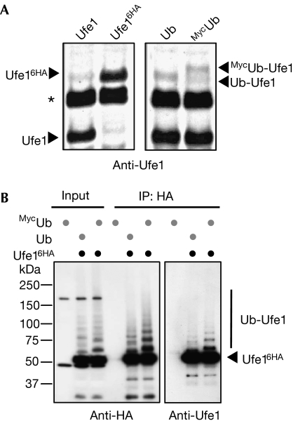
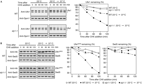
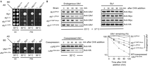
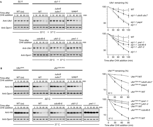
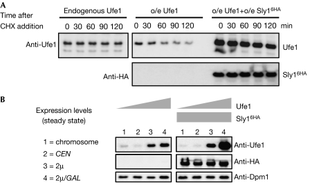
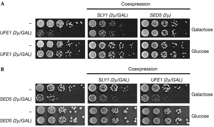
Endoplasmic reticulum (ER)-associated degradation (ERAD) is a specialized activity of the ubiquitin-proteasome system that is involved in clearing the ER of aberrant proteins and regulating the levels of specific ER-resident proteins. Here we show that the yeast ER-SNARE Ufe1, a syntaxin (Qa-SNARE) involved in ER membrane fusion and retrograde transport from the Golgi to the ER, is prone to degradation by an ERAD-like mechanism. Notably, Ufe1 is protected against degradation through binding to Sly1, a known SNARE regulator of the Sec1-Munc18 (SM) protein family. This mechanism is specific for Ufe1, as the stability of another Sly1 partner, the Golgi Qa-SNARE Sed5, is not influenced by Sly1 interaction. Thus, our findings identify Sly1 as a discriminating regulator of SNARE levels and indicate that Sly1-controlled ERAD might regulate the balance between different Qa-SNARE proteins.


Figures

Figure 1
Figure 1
The ER-SNARE Ufe1 is ubiquitylated in vivo. (A) Anti-Ufe1 immunoblots of extracts from cells expressing Ufe1 or a carboxy-terminally epitope-tagged Ufe1 form (Ufe16HA; left panel), or from cells coexpressing ubiquitin or an amino-terminally epitope-tagged ubiquitin (MycUb; right panel). UFE1 (UFE16HA) was expressed from its chromosomal locus. The positions of ubiquitin- (Ub-) and MycUb-modified Ufe1 forms are indicated by arrowheads. The asterisk denotes an unrelated crossreacting protein. (B) Immunoprecipitation of C-terminally HA-tagged Ufe1 (Ufe16HA) under denaturing conditions. WT and tagged Ufe1 proteins were expressed in Δufe1 cells at endogenous levels in combination with overexpressed Ub or MycUb. The input and the precipitated material were analysed by western blots using HA- (left panel) or Ufe1-specific antibodies (right panel). The positions of unmodified Ufe1 (Ufe16HA) and ubiquitin-modified Ufe1 forms (Ub-Ufe1) are indicated. ER, endoplasmic reticulum; HA, haemagglutinin; Ub, ubiquitin; WT, wild type.
Figure 2
Figure 2
Ufe1 but not Sed5 is unstable in the sly1-1 mutant. (A) CHX-chase experiment with WT (upper panel) and sly1-1 cells (lower panel). Cells were grown in YPD to an OD600 of 0.5 at 23°C and maintained at this temperature (left) or shifted up to 37°C after the addition of CHX (right). At each time point, the cellular level of Ufe1 was analysed by anti-Ufe1 immunoblots. As a control, the blots were re-probed with an antibody against the stable ER membrane protein Dpm1. The asterisk indicates a crossreactive band. The graph shows the quantification of the immunoblot signals from the Ufe1 decay (time point zero was set as 100%). (B) Similar to (A) but with strains expressing HA-tagged Ufe1 (Ufe16HA). Extracts were probed with HA (for tagged Ufe1), Sed5 or Dpm1 antibodies. CHX, cycloheximide; ER, endoplasmic reticulum; HA, haemagglutinin; OD, optical density; WT, wild type.
Figure 3
Figure 3
Loss of Ufe1–Sly1 interaction results in degradation of Ufe1. (A) Two-hybrid interactions between WT or mutant forms of Sly1 and Ufe1 fused to the activating domain (AD) or DNA-binding domain (BD) of Gal4. Transformants were spotted onto control (co) or media plates without histidine (−his) and incubated for 2–3 days. (B) Upper panel: CHX-chase experiments with Δsly1 cells expressing epitope-tagged proteins of either WT SLY1 or different sly1 mutants at endogenous level. The experimental set-up was essentially as described in Fig 2. Protein stability of Ufe1 and the respective Sly1 variant was monitored by anti-Ufe1 (left) and anti-Myc- or anti-HA immunoblots (right), respectively. Lower left panel: anti-HA immunoblot of CHX chase performed at 30°C with WT cells coexpressing plasmid-borne copies of HA-tagged variants of WT UFE1 or ufe1-F9A at endogenous level. The graph (lower right) shows quantified levels of endogenous Ufe1 and coexpressed Ufe1-F9A6HA in the respective strain backgrounds as indicated. CHX, cycloheximide; HA, haemagglutinin; WT, wild type.
Figure 4
Figure 4
Destabilized Ufe1 is degraded by an ERAD-like pathway. (A) Anti-Ufe1 immunoblots of CHX-chase experiments using WT (control; co) and sly1-1 mutant cells in which additional mutations (Δubc6 Δubc7, Δpep4, cdc48-6, ufd1-2, pre1-1) were introduced. The experimental set-up was essentially as in Fig 2. Representative experiments are shown and the graphs present the quantification of the Ufe1 decay resulting from 3 to 4 independent data sets; symbols and bars represent mean and s.e. (B) Anti-HA immunoblots of a CHX-chase experiment using WT (control; co) and mutants (same as above) coexpressing HA-tagged proteins of WT UFE1 or ufe1-F9A at endogenous UFE1 levels. The experimental procedure was similar to (A) except that the strains shown in the upper panel were grown to an OD600 of 0.6 at 30°C and kept at this temperature during the time course, whereas the strains shown in the lower panel were grown to an OD600 of 0.4 at 23°C and then shifted for an additional 2 h to 37°C before the CHX chase was started. Graphs: mean and s.e. of 3–4 independent data sets (Δpep4, n=2; error bars are deviation from the mean). CHX, cycloheximide; HA, haemagglutinin; OD, optical density; WT, wild type.
Figure 5
Figure 5
The ratio between Ufe1 and Sly1 affects Ufe1 stability. (A) Expression shut-off experiment monitoring Ufe1 protein stability when expressed endogenously (left panel) or on overexpression (o/e; right panel) in the absence or presence of additionally overexpressed Sly16HA. In case of overexpression, less sample was subjected to SDS-polyacrylamide gel electrophoresis to visualize protein decay. (B) Steady-state levels of Ufe1 detected by anti-Ufe1 immunoblots (left panel). UFE1 was expressed from (1) its genomic locus, (2) a low-copy-number centromeric plasmid, or (3) a high-copy-number 2μ-based plasmid, in each case driven by the UFE1 promoter, or (4) from a 2μ-based plasmid driven by the GAL1-10 promoter. The panels on the right show the same experimental design except that Sly1 (Sly16HA) is co-overexpressed from a 2μ-based plasmid under the control of the GAL1-10 promoter. Dpm1 levels were used as control. HA, haemagglutinin.
Figure 6
Figure 6
The ratio between Ufe1, Sed5 and Sly1 affects cellular growth. (A) Cytotoxicity mediated by UFE1 overexpression is reverted by concomitant overexpression of SLY1 or SED5. Cells overexpressing UFE1 alone (left), or together with SLY1 (middle), or SED5 (right) were adapted to galactose-containing media and then plated in fivefold dilutions on media containing galactose or glucose (as control) and incubated for 2–3 days at 30°C. All constructs were expressed in the DF5 strain from 2μ-based high-copy-number plasmids under pGAL1-10 control (except SED5: endogenous promoter). Empty vectors are denoted as ‘−'. (B) Cytotoxicity mediated by SED5 overexpression is partly reverted by concomitant overexpression of SLY1 or UFE1. Cells overexpressing SED5 alone (left), or together with SLY1 (middle), or UFE1 (right) were grown in liquid synthetic media containing raffinose and then plated in fivefold dilutions on media containing galactose or glucose (as control) and incubated for 2–3 days at 30°C. All constructs were expressed in the W303 strain from high-copy-number plasmids under pGAL1-10 control.

References

    1. Biederer T, Volkwein C, Sommer T (1997) Role of Cue1p in ubiquitination and degradation at the ER surface. Science 278: 1806–1809 - PubMed
    1. Bracher A, Weissenhorn W (2002) Structural basis for the Golgi membrane recruitment of Sly1p by Sed5p. EMBO J 21: 6114–6124 - PMC - PubMed
    1. Braun S, Matuschewski K, Rape M, Thoms S, Jentsch S (2002) Role of the ubiquitin-selective CDC48(UFD1/NPL4) chaperone (segregase) in ERAD of OLE1 and other substrates. EMBO J 21: 615–621 - PMC - PubMed
    1. Bryant NJ, James DE (2001) Vps45p stabilizes the syntaxin homologue Tlg2p and positively regulates SNARE complex formation. EMBO J 20: 3380–3388 - PMC - PubMed
    1. Cao X, Ballew N, Barlowe C (1998) Initial docking of ER-derived vesicles requires Uso1p and Ypt1p but is independent of SNARE proteins. EMBO J 17: 2156–2165 - PMC - PubMed

Publication types

MeSH terms