. 2007 Nov;115(11):1631-7.
doi: 10.1289/ehp.10216.

Nanosize titanium dioxide stimulates reactive oxygen species in brain microglia and damages neurons in vitro

Affiliations

Nanosize titanium dioxide stimulates reactive oxygen species in brain microglia and damages neurons in vitro

Thomas C Long et al. Environ Health Perspect. 2007 Nov.

Abstract

Background: Titanium dioxide is a widely used nanomaterial whose photo-reactivity suggests that it could damage biological targets (e.g., brain) through oxidative stress (OS).

Objectives: Brain cultures of immortalized mouse microglia (BV2), rat dopaminergic (DA) neurons (N27), and primary cultures of embryonic rat striatum, were exposed to Degussa P25, a commercially available TiO(2) nanomaterial. Physical properties of P25 were measured under conditions that paralleled biological measures.

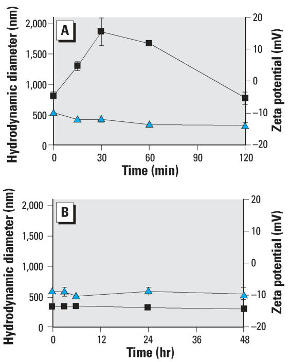
Findings: P25 rapidly aggregated in physiological buffer (800-1,900 nm; 25 degrees C) and exposure media (approximately 330 nm; 37 degrees C), and maintained a negative zeta potential in both buffer (-12.2 +/- 1.6 mV) and media (-9.1 +/- 1.2 mV). BV2 microglia exposed to P25 (2.5-120 ppm) responded with an immediate and prolonged release of reactive oxygen species (ROS). Hoechst nuclear stain was reduced after 24-hr (>or=100 ppm) and 48-hr (>or=2.5 ppm) exposure. Microarray analysis on P25-exposed BV2 microglia indicated up-regulation of inflammatory, apoptotic, and cell cycling pathways and down-regulation of energy metabolism. P25 (2.5-120 ppm) stimulated increases of intracellular ATP and caspase 3/7 activity in isolated N27 neurons (24-48 hr) but did not produce cytotoxicity after 72-hr exposure. Primary cultures of rat striatum exposed to P25 (5 ppm) showed a reduction of immunohistochemically stained neurons and microscopic evidence of neuronal apoptosis after 6-hr exposure. These findings indicate that P25 stimulates ROS in BV2 microglia and is nontoxic to isolated N27 neurons. However, P25 rapidly damages neurons at low concentrations in complex brain cultures, plausibly though microglial generated ROS.

Keywords: BV2; P25; environmental nanotoxicity; neurotoxicity; oxidative stress; titanium dioxide.


Figures

Figure 1
Figure 1
(A) The immediate production of intracellular H2O2 generated from the oxidative burst was measured in BV2 microglia with Image-iT LIVE Green. Cells were incubated (30 min, 37°C) in 25 μM Image-iT LIVE Green and exposed to P25. Significant increases of fluorescence first occurred in response to P25 (60 ppm; 1 min). (B) The production of formula image resulting from interference with the mitochondria’s ETC was measured with MitoSOX Red. BV2 microglia, incubated in 2 μM (10 min, 37°C) showed a delayed but significant increase in fluorescence after 30-min exposure to ≥100 ppm P25. (C) Significant increases of caspase 3/7 activity were first seen by 6 hr in response to ≥40 ppm P25 and remained at this level for 24 hr. (D) Apoptotic loss of nuclear material, as measured with Hoechst stain, was first noted after 24 hr in response to P25 (≥100 ppm) and involved all concentrations by 48 hr.
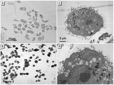
Figure 2
Figure 2
BV2 microglia exposed to P25 (20 ppm) were examined with both LM and TEM. (A) The toluidine blue stained cytoplasm of BV2 microglia housed numerous, light-refractive P25 aggregates after 3-hr exposure. (B) LM examination of unstained, fixed cells exposed to P25 for 48 hr indicated that the cellular membranes were fragmented and showed granular cytoplasm and centralized nuclei. Magnification × 1,200. (C) TEM examination of the P25 exposed BV2 microglia indicate phagocytic internalization of the P25 aggregates after 3 hr. (D) Higher magnification of the BV2 microglial cytoplasm indicated swollen and disrupted mitochondria (circles) in proximity to the P25 aggregates.
Figure 3
Figure 3
BV2 microglia were exposed to P25 (20 ppm) for 3 hr and prepared for microarray analysis. IL-4, interleukin 4; PPARα, peroxisome proliferator-activated receptor α (A) IPA’s Core analysis (metabolic/signaling pathways) indicated that up-regulated genes were clustered around signaling pathways involved with apoptosis, Death receptor families (i.e., caspase activation), calcium signaling, inflammation (NF-κB), and cell cycling and maintenance. (B) Toxicity Pathway analysis indicated that P25 up-regulated pathways were primarily associated with inflammatory (NF-κB), cell cycling and pro-apoptotic activities.
Figure 4
Figure 4
BV2 microglia were exposed to P25 (20 ppm) for 3 hr. IL-6, interleukin 6, PPARα, peroxisome proliferator-activated receptor α TR/RXR, thyroid hormone receptor/retinoid X receptor. (A) Core analysis of the down-regulated genes showed clustering around pathways associated with adaptive change and key energy production pathways. (B) Toxicity Pathway analysis indicated that P25 down-regulated genes in pathways associated with hypoxia, peroxisomes, and Nrf2-mediated oxidative stress.
Figure 5
Figure 5
BV2 microglia were exposed to P25 (20 ppm) for 3 hr. (A) Canonical analysis of all P25 affected genes associated with OS indicated that they largely clustered around key energy pathways involving oxidative phosphorylation, biosynthesis of ubiquinone (involved in shuttling electrons in the ETC) and the citric acid cycle. (B) Toxicity Pathway analysis localized these pathways further to mitochondrial dysfunction.
Figure 6
Figure 6
(A) N27 neurons were exposed (1–72 hr) to P25 (2.5–120 ppm) and intracellular ATP levels measured with CellTiter-Glo. Significant increases were seen as early as 1 hr postexposure to ≥80 ppm and continued until 48 hr in response to ≥40 ppm. (B) Significant increases (p < 0.05) in caspase 3/7 activity were first seen in N27 neurons after 24 hr in response to ≥40 ppm P25. (C) Significant reductions of Hoechst stain did not occur in response to P25 (2.5–120 ppm) at any time point. (D) TEM of P25 (20 ppm, 3 hr) treated N27 neurons showed numerous membrane-bound aggregates. An amorphous substance was seen within the vacuoles (insert). In addition, individual nanosize P25 particles (circle) were noted throughout the cytoplasm. Mitochondria in nearby proximity showed no evidence of disruption or swelling.
Figure 7
Figure 7
LM histology of IHC rat embryonic striatum. Confluent cultures of embryonic were exposed to 5 ppm P25 for 6–48 hr, IHC stained with NSE and morphometrically analyzed. (A) Untreated cultures consisted of a dense plexus of neurons and glia. (B) Axonal beading and cellular granularity were seen as early as 6 hr postexposure. (C) Evidence of apoptosis (circles) was documented by 24 hr. (D) Complete disruption and loss of cellular integrity was noted by 48 hr postexposure to 5 ppm P25.
Figure 8
Figure 8
Morphometric analysis was conducted on NSE-stained cultures of mouse striatum. These data indicated that the total area of NSE-stained neurons was reduced by 14% after 6-hr exposure (A) and 19% after 24 hr (B) to P25 (5 ppm).
Figure 9
Figure 9
The aggregate size and zeta potential of P25 (20 ppm) were measured in both HBSS and RPMI at conditions that paralleled the biological responses. (A) P25 aggregates reached 1,900 nm in size over a 30-min measurement in HBSS (25°C) and maintained > 1,000 nm size for the 2-hr exposure period. The zeta potential of P25 (blue triangles) initially measured –9.8 mV and decreased slightly over the 2-hr period. (B) Both the aggregate size (black squares; 300–350 nm) and zeta potential (blue triangles) (–8 mV to –10 mV) of P25 remained stable when measured in RPMI over the 48-hr period.

References

    1. Adams LK, Lyon DY, McIntosh A, Alvarez PJ. Comparative toxicity of nano-scale TiO2, SiO2 and ZnO water suspensions. Water Sci Technol. 2006;54:327–334. - PubMed
    1. Afaq F, Abidi P, Matin R, Rahman Q. Cytotoxicity, pro-oxidant effects and antioxidant depletion in rat lung alveolar macrophages exposed to ultrafine titanium dioxide. J Appl Toxicol. 1998;18:307–312. - PubMed
    1. Affymetrix Inc. Affymetrix Technical Manual. 2004. [[accessed 2 October 2007]]. Available: www.expressionanalysis.comm.
    1. Artuch R, Colome C, Vilaseca MA, Pineda M, Campistol J. Ubiquinone: metabolism and functions. Ubiquinone deficiency and its implication in mitochondrial encephalopathies. Treatment with ubiquinone. Rev Neurol. 1999;29:59–63. - PubMed
    1. Beck-Speier I, Dayal N, Karg E, Maier KL, Roth C, Ziesenis A, et al. Agglomerates of ultrafine particles of elemental carbon and TiO2 induce generation of lipid mediators in alveolar macrophages. Environ Health Perspect. 2001;109(suppl 4):613–618. - PMC - PubMed

MeSH terms

LinkOut - more resources