. 2007 Dec;117(12):3765-73.
doi: 10.1172/JCI32538.

FOXP3 is a novel transcriptional repressor for the breast cancer oncogene SKP2

Affiliations

FOXP3 is a novel transcriptional repressor for the breast cancer oncogene SKP2

Tao Zuo et al. J Clin Invest. 2007 Dec.

Abstract

S-phase kinase-associated protein 2 (SKP2) is a component of the E3 ubiquitin ligase SKP1-Cul1-Fbox complex. Overexpression of SKP2 results in cell cycle dysregulation and carcinogenesis; however, the genetic lesions that cause this upregulation are poorly understood. We recently demonstrated that forkhead box P3 (FOXP3) is an X-linked breast cancer suppressor and an important repressor of the oncogene ERBB2/HER2. Since FOXP3 suppresses tumor growth regardless of whether the tumors overexpress ERBB2/HER2, additional FOXP3 targets may be involved in its tumor suppressor activity. Here, we show that mammary carcinomas from mice heterozygous for a Foxp3 mutation exhibited increased Skp2 expression. Ectopic expression of FOXP3 in mouse mammary cancer cells repressed SKP2 expression with a corresponding increase in p27 and polyploidy. Conversely, siRNA silencing of the FOXP3 gene in human mammary epithelial cells increased SKP2 expression. We also show that Foxp3 directly interacted with and repressed the Skp2 promoter. Moreover, the analysis of over 200 primary breast cancer samples revealed an inverse correlation between FOXP3 and SKP2 levels. Finally, we demonstrated that downregulation of SKP2 was critical for FOXP3-mediated growth inhibition in breast cancer cells that do not overexpress ERBB2/HER2. Our data provide genetic, biochemical, and functional evidence that FOXP3 is a novel transcriptional repressor for the oncogene SKP2.


Figures
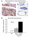
Figure 1
Figure 1. A naturally occurring mutation in the Foxp3 locus results in increased Skp2 expression.
(A) Immunohistochemical staining of the Skp2 protein in mammary cancers and an adjacent normal mammary gland in 1 Foxp3sf/+ mouse. Original magnification, ×40. (B) Relative levels of Skp2 transcripts in normal mammary epithelium of WT and Foxp3sf/+ mice and the cancerous tissue in the Foxp3sf/+ mice, as revealed by real-time RT-PCR of LCM samples. The expression of Skp2 was normalized against the internal control, the Hprt gene. Data shown are means ± SD of 3 samples in each group. Highly significant differences were observed between cancerous and normal tissue (P < 0.001, ANOVA test when either internal standards were used).
Figure 2
Figure 2. Foxp3 represses Skp2 transcription.
(A) Transfection of Foxp3-V5 into TSA cells repressed expression of the Skp2 gene. The mRNA levels of Foxp3, Skp2, and p27 were measured by real-time PCR for the vector control cells, polyclonal Foxp3-V5 transfectants (CL30), and 2 stable Foxp3-V5 transfectant clones, CL302 and CL305. Data shown are relative amounts of transcripts after normalizing against the amounts of total RNA based on the levels of Hprt mRNA. The means of the vector group are artificially defined as 1.0. Data shown are means ± SD of 3 independent experiments. (B) Foxp3 reduces Skp2 with a corresponding increase in p27. Lysates of Foxp3-V5 or vector-transfected TSA cell lines were analyzed by Western blot using SKP2, p27, β-actin, and anti-V5 (which recognize V5-tagged Foxp3) antibodies. (C) Foxp3 increased stability of p27. Vector or Foxp3-V5 transfectants were treated with cycloheximide (CHX, 100 μM) for the indicated intervals. Cells were collected and p27 protein levels were detected by Western blot. The upper panel shows representative experiments while the lower panel shows the decay of p27, using time 0 as 1.0. Protein loading equivalence was assessed by the expression of β-actin. The relative intensity of bands was measured relative to their respective β-actin bands using BandScan software (version 4.3; Glyco). (D) Polyubiquitination of the p27 in vector or Foxp3 transfectants. Vector or Foxp3-V5–transfected TSA cell lines were either left untreated or treated with proteasome inhibitor MG132 (40 μM) for 3 hours. Equal aliquot of the cellular lysates were immunoprecipitated with anti-p27 antibodies. The precipitates were separated by SDS-PAGE and transferred into nitrocellulose membrane, which were then autoclaved in water for 30 minutes and blotted with HRP-conjugated anti-polyubiquitin antibody Ub P4D1 (33).
Figure 3
Figure 3. Foxp3 binding to Skp2 is important for transcriptional repression.
(A) Foxp3 represses mouse Skp2 promoter activity. Either Foxp3 cDNA or empty vector was transiently cotransfected with reporter vector at different ratios illustrated in the figure. Cells were transfected with either vector control or Foxp3 (1 μg/well) in conjunction with the luciferase reporter driven by 5′ promoter regions of the Skp2 gene (0.2 μg, 0.4 μg and 1.0 μg per well). Forty-eight hours later, the cell lysates were harvested and measured for luciferase activity. The luciferase activity from the cells transfected with the pGL2-basic vector was arbitrarily defined as 1.0. Data shown are means ± SD of triplicates and have been repeated at least 3 times. (B) The upper panel depicts the 5′ region of the Skp2 gene. The lower panel shows the amount of DNA precipitated by anti-V5 mAbs after subtracting a minute portion precipitated by IgG control. The data shown are a fraction of the total genomic DNA isolated from the same number of cells. (C) Deletion of 1 of the 2 Foxp3-binding sites in the Skp2 promoter region prevented FOXP3-mediated suppression. The deleted sequences are mut A: ACTAAACCAATATTCTAAT and mut B: TAAAAATAAACCATC. The promoter activity was measured in the human breast cancer line T47D.
Figure 4
Figure 4. Polyploidy of the Foxp3hi but not the Foxp3lo mouse mammary cancer cells.
(A) Modest Foxp3-V5 expression in the Foxp3 transfectant compared with either untransfected parental cells or control vector-transfected cells. Data shown in the upper panel are histograms depicting Foxp3-V5 protein levels, while those in the lower panel show levels of FoxP3 mRNA in Foxp3-V5- or vecotr-transfected TSA or LCM captured mouse mammary epithelial cells (MME), as measured by quantitative real-time PCR. (B) Foxp3 expression caused polyploidy. Left panel shows the level of the Foxp3-V5 fusion protein in vector control (top) or Foxp3-transfected TSA cells (bottom) while the right panel shows the DNA contents of vector control (top), Foxp3hi (middle), and Foxp3lo cells (bottom). (C) Ectopic expression of Skp2 alleviated polyploidy induced by Foxp3. Vector (i, ii) or Foxp3 (iii, iv) transfectants of TSA were transfected with either vector control or Skp2. After removing untransfected cells with blasticidin, the transfectants were fixed with Cytofix/Cytoperm buffer (BD) and tested with DNA contents using 7-AAD. Representative profiles are shown in the left and middle panels while summary data from 3 independent experiments are shown in the right panel. *P < 0.05.
Figure 5
Figure 5. Silencing of FOXP3 resulted in upregulation of SKP2 in primary HMEC.
FOXP3 was silenced in HMEC by using siRNA. FOXP3 and SKP2 transcripts were quantified by real-time PCR. The RNA inputs were normalized against housekeeping gene GAPDH. Data shown are means ± SEM of relative levels of FOXP3 and represent 3 independent experiments (P < 0.01, Student’s t test). In A, the vector control was defined as 1.0; in B, silenced cells were defined as 1.0. Data shown are means ± SD of triplicates and represent 3 independent experiments.
Figure 6
Figure 6. Inducible FOXP3 expression in TetOff breast cancer cells rapidly downregulated SKP2 expression.
Upper panel shows a diagram depicting the promoter structure in pBI-EGFP vector. The bidirectional promoter Pbi-1 was responsive to the tTA regulatory proteins in the TetOff system. Pbi-1 contains the tetracycline-responsive element (TRE) that is between 2 identical minimal CMV promoters (PminCMV). EGFP is at one side while human FOXP3 cDNA was inserted at the PvuII/NheI sites in the opposite direction. A control vector with GFP but not FOXP3 cDNA was also used. Lower panel presents kinetics of SKP2 repression by FOXP3. FOXP3 was induced by the withdrawal of deoxycycline in medium. The transcript levels were first normalized for RNA input by using GAPDH and then compared with those observed in the control cell lines, defined as 1.0.
Figure 7
Figure 7. Significant reduction in the rate of SKP2 upregulation in the FOXP3+ breast cancer samples.
Tissue microarray samples of human breast cancer were stained with either anti-FOXP3 antibody or anti-SKP2 antibody. The samples were scored in a double-blind fashion. The top panels show staining patterns of either FOXP3 or SKP2 in 2 representative cases. Original magnification, ×60. Summary data from 206 independent cases are presented in the lower panel. The P values of the χ2 tests are listed.
Figure 8
Figure 8. Ectopic expression of Skp2 substantially alleviates growth inhibition of MCF-7 cell lines by FOXP3.
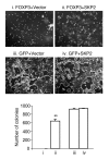
MCF-7 cell lines with inducible expression of either GFP (iii, iv) or FOXP3 (i, ii) were supertransfected with either vector control (i, iii) or SKP2 (ii, iv). After removing untransfected cells by drug selection, the cultures were maintained in tetracycline-free conditions for 2 weeks. Upper and middle panels show photographs of viable (ii, iii, iv) or apoptotic MCF-7 cells. Original magnification, ×10. The lower panel shows colony numbers per 100-mm2 plate. At the end of 2 weeks of culture, the dead cells were removed and the plates were stained with violet crystal. The visible colonies were counted. Data shown are means of SD of triplicates and are representative of 3 independent experiments. **P < 0.001 when group I and II were compared by Student’s t test.

References

    1. Huang H., et al. Skp2 inhibits FOXO1 in tumor suppression through ubiquitin-mediated degradation. Proc. Natl. Acad. Sci. U. S. A. 2005;102:1649–1654. - PMC - PubMed
    1. Pagano M. Control of DNA synthesis and mitosis by the Skp2-p27-Cdk1/2 axis. Mol. Cell. 2004;14:414–416. - PubMed
    1. Nakayama K.I., Nakayama K. Ubiquitin ligases: cell-cycle control and cancer. Nat. Rev. Cancer. 2006;6:369–381. - PubMed
    1. Schmidt M., et al. Cell cycle inhibition by FoxO forkhead transcription factors involves downregulation of cyclin D. Mol. Cell. Biol. 2002;22:7842–7852. - PMC - PubMed
    1. Zuo T., et al. FOXP3 is an X-linked breast cancer suppressor gene and an important repressor of HER-2/ErbB2 oncogene. Cell. . 2007;129:1275–1286. - PMC - PubMed

Publication types

MeSH terms