. 2008 Apr;76(4):348-56.
doi: 10.1111/j.1432-0436.2007.00232.x. Epub 2007 Nov 15.

Complementary roles for histone deacetylases 1, 2, and 3 in differentiation of pluripotent stem cells

Affiliations

Complementary roles for histone deacetylases 1, 2, and 3 in differentiation of pluripotent stem cells

Glen W Humphrey et al. Differentiation. 2008 Apr.

Abstract

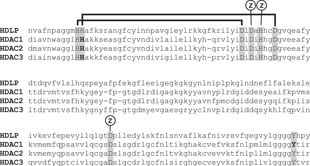
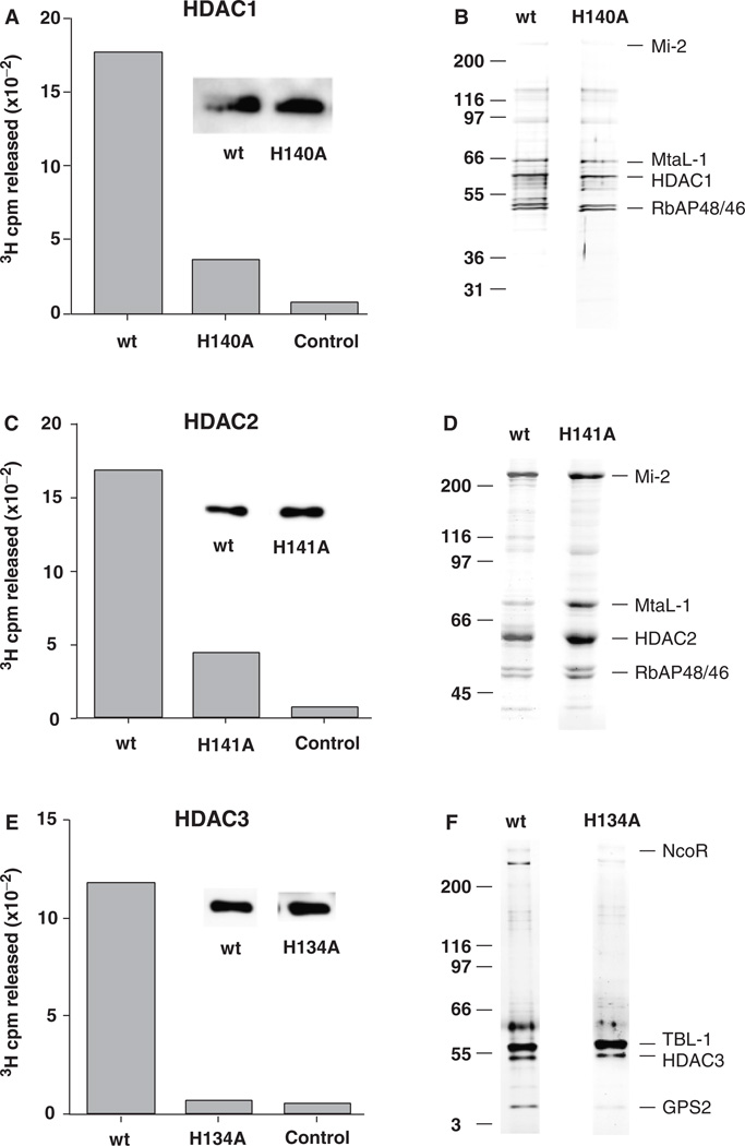
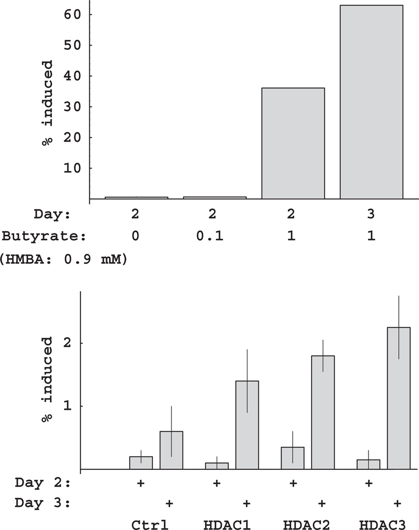
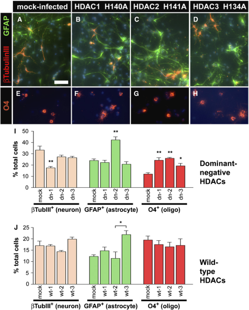
In eukaryotic cells, covalent modifications to core histones contribute to the establishment and maintenance of cellular phenotype via regulation of gene expression. Histone acetyltransferases (HATs) cooperate with histone deacetylases (HDACs) to establish and maintain specific patterns of histone acetylation. HDAC inhibitors can cause pluripotent stem cells to cease proliferating and enter terminal differentiation pathways in culture. To better define the roles of individual HDACs in stem cell differentiation, we have constructed "dominant-negative" stem cell lines expressing mutant, Flag-tagged HDACs with reduced enzymatic activity. Replacement of a single residue (His-->Ala) in the catalytic center reduced the activity of HDACs 1 and 2 by 80%, and abolished HDAC3 activity; the mutant HDACs were expressed at similar levels and in the same multiprotein complexes as wild-type HDACs. Hexamethylene bisacetamide-induced MEL cell differentiation was potentiated by the individual mutant HDACs, but only to 2%, versus 60% for an HDAC inhibitor, sodium butyrate, suggesting that inhibition of multiple HDACs is required for full potentiation. Cultured E14.5 cortical stem cells differentiate to neurons, astrocytes, and oligodendrocytes upon withdrawal of basic fibroblast growth factor. Transduction of stem cells with mutant HDACs 1, 2, or 3 shifted cell fate choice toward oligodendrocytes. Mutant HDAC2 also increased differentiation to astrocytes, while mutant HDAC1 reduced differentiation to neurons by 50%. These results indicate that HDAC activity inhibits differentiation to oligodendrocytes, and that HDAC2 activity specifically inhibits differentiation to astrocytes, while HDAC1 activity is required for differentiation to neurons.


Figures

Fig. 1
Fig. 1
Conserved catalytic domain of type I histone deacetylases. Sequence alignment of Aquifex aeolicus HDLP (1C3R_B) with human HDAC1 (Q13547), HDAC2 (Q92769), and HDAC3 (O15379). Residues located in the catalytic center are in caps. Residues mutated for this study are in bold. Zinc-binding residues are indicated by “Z.” Horizontal lines connect the two His–Asp charge relays (see text). HDAC, histone deacetylases; HDLP, HDAC-like protein.
Fig. 2
Fig. 2
Wild-type (wt) and mutant HDACs form similar multiprotein complexes. Flag-tagged HDACs were expressed in Hela cells and isolated using an anti-Flag immunoaffinity adsorbent. HDAC1 enzymatic activity was determined by 3H cpm released from 3H acetate-labeled histones in a 30-min incubation (see “Methods”). (A) Wt, mutant (H140A) HDAC1 complexes isolated from transduced Hela cells; control anti-Flag adsorbent using normal (untransduced) Hela cells. Insets show that similar amounts of Flag-tagged protein were used for each assay. (C) Wt or mutant (H141A) HDAC2 complexes isolated from transduced Hela cells, control anti-Flag adsorbent using normal (untransduced) Hela cells. (E) Wt or mutant (H134A) HDAC3 complexes isolated from transduced Hela cells, control anti- Flag adsorbent using normal (untransduced) Hela cells. Wt and mutant HDACs form similar multiprotein complexes. (B) Comparison of immunopurified Flag-tagged HDAC1 wt and mutant (H140A) multiprotein complexes resolved by SDS-PAGE and visualized by silver staining. Right, principal components; left, size markers. (D) Comparison of immunopurified Flag-tagged HDAC2 wt and mutant (H141A) multiprotein complexes. (F) Comparison of wt and mutant (H134A) HDAC3 form similar multiprotein complexes. HDAC, histone deacetylases.
Fig. 3
Fig. 3
Potentiation of HMBA-induced MEL differentiation by inhibition of HDAC activity. (A) Potentiation of HMBA-induced MEL differentiation by sodium butyrate. MEL cultures were treated with 0, 0.1, or 1.0 mM butyrate in the presence of 0.9 mM HMBA and assayed for differentiation on days 2 and 3. (B) MEL differentiation following retroviral transduction with HDAC1-3 mutants. MEL cultures were transduced with a retrovirus containing no insert (Ctrl), or dominant-negative HDAC constructs (HDAC1, 2, or 3). Following treatment with 0.9 mM HMBA, cultures were assayed for differentiation on days 2 and 3. HDAC, histone deacetylases; HMBA, Hexamethylene bisacetamide.
Fig. 4
Fig. 4
Loss of HDAC function causes changes in stem cell fate. Stem cells were cultured from E14.5 rat forebrain and infected with a retrovirus containing no insert (A, E) or dominant-negative HDAC constructs (B–D, F–H). Infected cells were purified based on the expression of an IL2R selectable marker, cultured in bFGF for 2 days, and then differentiated by bFGF withdrawal for 7 additional days. Cells were immunostained for β-III-Tubulin to mark neurons (A–D, red), GFAP to identify astrocytes (A–D, green) or O4 to mark oligodendrocytes (E–H, red) and DAPI to identify cell nuclei (blue). (I) Quantitation of differentiated cells after mock or dominant-negative construct infections of stem cells (mean SEM, n = 4–9). (J) Quantitation of differentiated cells after mock or wild-type construct infections of stem cells. CNPase is used instead of O4 to mark oligodendrocytes. Lower baseline values reflect slower differentiation due to lower initial plating densities (mean ± SEM, n = 2). *p < 0.05; **p < 0.001. Bar = 200 µm for all images. HDAC, histone deacetylases; bFGF, basic fibroblast growth factor; GFAP, glial fibrillary acidic protein.

References

    1. Ballas N, Battaglioli E, Atouf F, Andres ME, Chenoweth J, Anderson ME, Burger C, Moniwa M, Davie JR, Bowers WJ, Federoff HJ, Rose DW, Rosenfeld MG, Brehm P, Mandel G. Regulation of neuronal traits by a novel transcriptional complex. Neuron. 2001;31:353–365. - PubMed
    1. Borre A, Cultraro CM, Segal S. c-Myc inactivation by mutant Max alters growth and morphology of NCI-H-630 colon cancer cells. J Cell Physiol. 1996;169:200–208. - PubMed
    1. Brehm A, Miska EA, McCance DJ, Reid JL, Bannister AJ, Kouzarides T. Retinoblastoma protein recruits histone deacetylase to repress transcription. Nature. 1998;391:597–601. - PubMed
    1. Chuang DM. The antiapoptotic actions of mood stabilizers: molecular mechanisms and therapeutic potentials. Ann N Y Acad Sci. 2005;1053:195–204. - PubMed
    1. Fajas L, Egler V, Reiter R, Hansen J, Kristiansen K, Debril MB, Miard S, Auwerx J. The retinoblastoma-histone deacetylase 3 complex inhibits PPARgamma and adipocyte differentiation. Dev Cell. 2002;3:903–910. - PubMed

Publication types