. 2007 Nov 16:5:58.
doi: 10.1186/1479-5876-5-58.

N-myristoyltransferase: a potential novel diagnostic marker for colon cancer

Affiliations

N-myristoyltransferase: a potential novel diagnostic marker for colon cancer

Anuraag Shrivastav et al. J Transl Med. .

Abstract

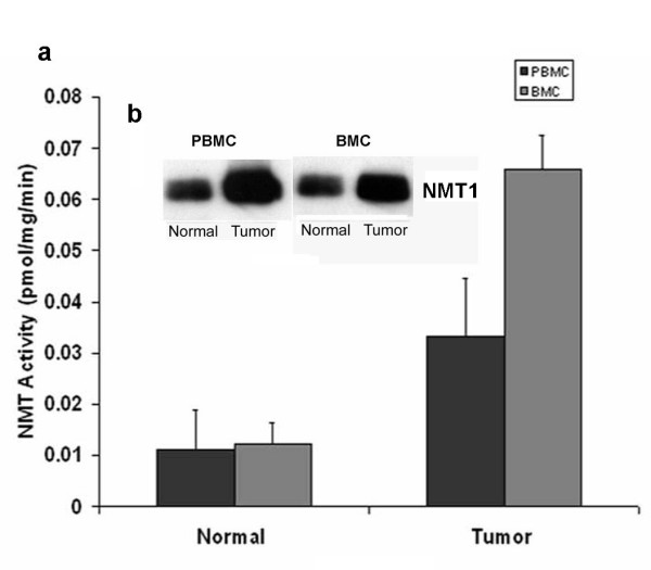
Background: Colon cancer is the second leading cause of cancer deaths in the western world. If detected early, colorectal cancer is one of the most treatable forms of cancer. Unfortunately, very few people are screened. N-myristoyltransferase (NMT) catalyzes myristoylation of various proteins including oncoproteins. We have demonstrated earlier the alteration of NMT activity during the progression of colorectal cancer and established NMT as a putative therapeutic target for cancer.

Methods: Peripheral blood samples and bone marrow were collected from the colon cancer patients and azoxymethane induced colonic tumor rats and their controls respectively. NMT activity and expression was determined as reported earlier. Immunohistochemical studies were carried out using standard procedures.

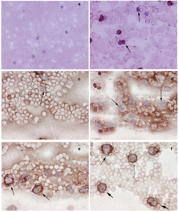
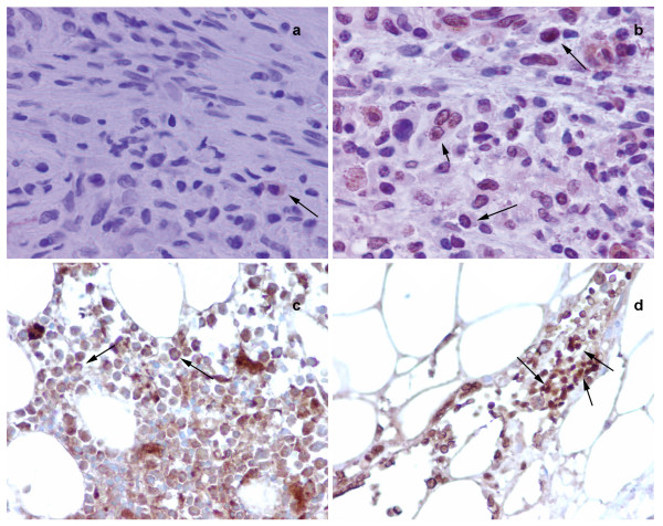
Results: In this study we demonstrate for the first time altered expression and localization of NMT in the peripheral blood and bone marrow in colon cancer patients. Immunohistochemical analysis revealed weak to negative staining for NMT in peripheral blood mononuclear cells (PBMC) of controls, whereas strong positivity was observed in PBMC colon cancer patients. In addition, we observed that NMT was localized mostly in the nuclei of the bone marrow (BM) mononuclear cells of the colon cancer patients, whereas NMT remained cytoplasmic in the control bone marrow specimens.

Conclusion: The strikingly different NMT expression offers the basis of a potential adjunct investigative tool for screening or diagnosis of patients at risk for or suspected of having colon cancer. Furthermore, altered localization of NMT in BM of tumor bearing hosts may serve as an added investigative tool for the diagnostic purpose.


Figures

Figure 1
Figure 1
NMT activity in peripheral blood mononuclear cells (PBMC) and Bone Marrow Cells (BMC) of normal and colorectal tumor bearing rats a; Isolated peripheral blood mononuclear cells from peripheral blood of control or tumor bearing rat were assessed for NMT activity. NMT activity was assayed using cAMP-dependent protein kinase derived peptide substrate. Values are mean ± SD of three independent experiments. Inset: Western blot analysis of peripheral blood mononuclear cells and bone marrow cells of normal and colorectal tumor bearing rats. Proteins (25 μg) from PBMC or BMC of control or tumor bearing rats were subjected to 10% SDS-PAGE, transblotted onto nitrocellulose membrane and were probed with monoclonal anti-human NMT antibody (1:250 dilutions).
Figure 2
Figure 2
Immunohistochemical analysis of peripheral blood mononuclear cells (PBMC) of normal and tumor bearing hosts. Smears of peripheral blood cells were incubated with anti-NMT antibody. Peripheral blood mononuclear cells (mostly lymphocytes) from control rats were devoid of NMT staining (a), whereas, intense NMT expression was observed in the peripheral blood mononuclear cells of colorectal tumor bearing rats as evident from strong staining (b, see arrows). c negative staining of lymphocytes (see arrow) & d; shows negative staining of monocytes (see arrows) in peripheral blood smear of control e; peripheral blood smear of colon cancer patients show positive staining of macrophages (arrows), f; peripheral blood smear of colon cancer patients show positive staining of neutrophil (fat arrows), lymphocyte (lean long arrow) and macrophages (arrow).
Figure 3
Figure 3
Immunohistochemical analysis of Bone Marrow of normal and tumor bearing hosts. a; shows cytoplasmic staining of NMT (see arrow) in bone marrow cells from control rats, b; whereas, most of the cells shows nuclear localization of NMT in bone marrow cells from tumor bearing rat (see arrow). c; NMT staining is mostly cytoplasmic in bone marrow of control (see arrow). d; Intense nuclear (and some cytoplasmic) staining for NMT is observed in the bone marrow of colon cancer patient (see arrow).

References

    1. Boutin JA. Myristoylation. Cell Signal. 1997;9:15–35. doi: 10.1016/S0898-6568(96)00100-3. - DOI - PubMed
    1. Resh MD. Fatty acylation of proteins: new insights into membrane targeting of myristoylated and palmitoylated proteins. Biochim Biophys Acta. 1999;1451:1–16. doi: 10.1016/S0167-4889(99)00075-0. - DOI - PubMed
    1. Farazi TA, Waksman G, Gordon JI. The biology and enzymology of protein N-myristoylation. J Biol Chem. 2001;276:39501–39504. doi: 10.1074/jbc.R100042200. - DOI - PubMed
    1. Rajala RV, Datla RS, Moyana TN, Kakkar R, Carlsen SA, Sharma RK. N-myristoyltransferase. Mol Cell Biochem. 2000;204:135–155. doi: 10.1023/A:1007012622030. - DOI - PubMed
    1. Shrivastav A, Selvakumar P, Bajaj G, Lu Y, Dimmock JR, Sharma RK. Regulation of N-myristoyltransferase by novel inhibitor proteins. Cell Biochem Biophys. 2005;43:189–202. doi: 10.1385/CBB:43:1:189. - DOI - PubMed

Publication types

LinkOut - more resources