. 2007 Nov 16:7:230.
doi: 10.1186/1471-2148-7-230.

The [FeFe] hydrogenase of Nyctotherus ovalis has a chimeric origin

Affiliations

The [FeFe] hydrogenase of Nyctotherus ovalis has a chimeric origin

Brigitte Boxma et al. BMC Evol Biol. .

Abstract

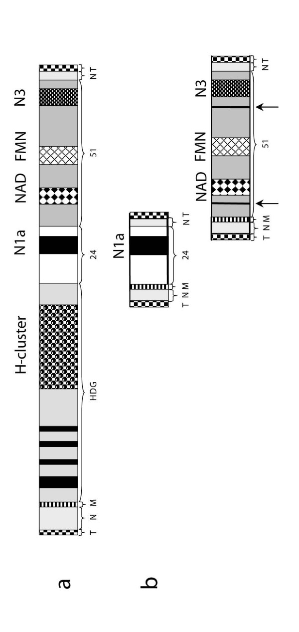
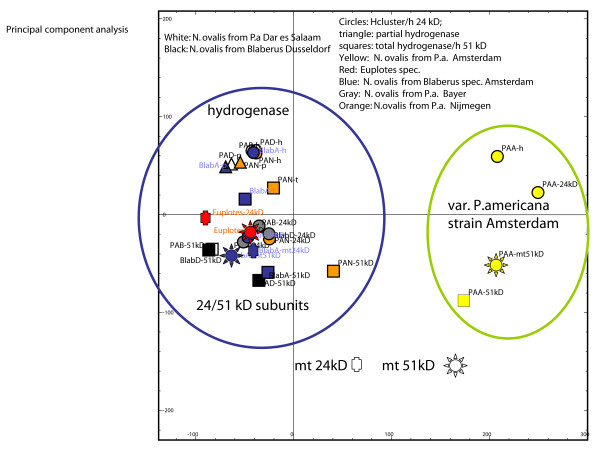
Background: The hydrogenosomes of the anaerobic ciliate Nyctotherus ovalis show how mitochondria can evolve into hydrogenosomes because they possess a mitochondrial genome and parts of an electron-transport chain on the one hand, and a hydrogenase on the other hand. The hydrogenase permits direct reoxidation of NADH because it consists of a [FeFe] hydrogenase module that is fused to two modules, which are homologous to the 24 kDa and the 51 kDa subunits of a mitochondrial complex I.

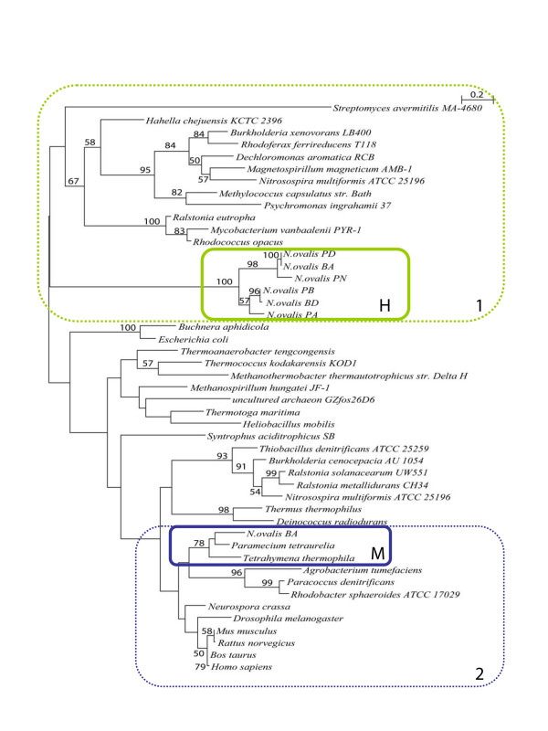
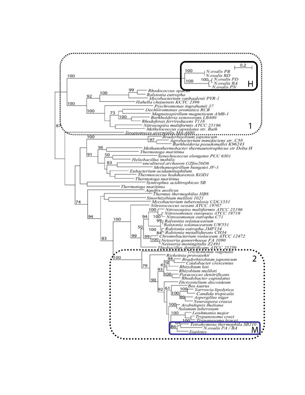
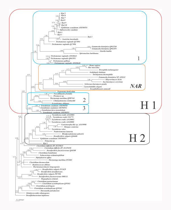
Results: The [FeFe] hydrogenase belongs to a clade of hydrogenases that are different from well-known eukaryotic hydrogenases. The 24 kDa and the 51 kDa modules are most closely related to homologous modules that function in bacterial [NiFe] hydrogenases. Paralogous, mitochondrial 24 kDa and 51 kDa modules function in the mitochondrial complex I in N. ovalis. The different hydrogenase modules have been fused to form a polyprotein that is targeted into the hydrogenosome.

Conclusion: The hydrogenase and their associated modules have most likely been acquired by independent lateral gene transfer from different sources. This scenario for a concerted lateral gene transfer is in agreement with the evolution of the hydrogenosome from a genuine ciliate mitochondrion by evolutionary tinkering.


Figures

Figure 1
Figure 1
Schematic representation of the minichromosomes encoding the hydrogenase (a) and the "mitochondrial" 24 and 51 kDa genes (b). The macronuclear minichromosomes are capped by telomeres (T) and contain non-coding DNA sequences (N) at the N- and C-terminal parts of the chromosome. A mitochondrial targeting signal (M) is found at the N terminal part of the coding sequence. 1. a. The hydrogenase is chimeric, i.e. it consists of a long-type [FeFe] hydrogenase with 4 FeS clusters (black bars in HDG), a 24 kDa (hoxF) module ("24") with an N1a type FeS cluster, and a 51 kDa (hoxU) ("51") module with a N3-type [4Fe-4S] cluster plus a FMN and a NAD binding site. 1. b. The subunits of the "mitochondrial" complex I are localized on individual minichromosomes. They each possess a mitochondrial targeting signal (M) and upstream and downstream non-coding DNA (N). The "mitochondrial" 51 kDa module possesses two small introns (arrows) that are absent from the correspondent hydrogenase module.
Figure 2
Figure 2
Phylogenetic tree of the 24 kDa-like module of the hydrogenase of N. ovalis, mitochondrial complex I 24 kDa subunits, bacterial NuoE, and bacterial hydrogenase subunits. See methods for the Accession Numbers and how the tree was calculated. H: N. ovalis hydrogenase, M: ciliate mitochondrial. Bootstraps are only indicated in the tree if they are ≥ 50. Box 1 marks 24 kDa modules that are fused with their corresponding 51 kDa modules (with the exception of Nitrosospira multiformis). All bacteria in this box (with the exception of Nitrosospira multiformis) have a [NiFe] hydrogenase. The mitochondrial/alpha-proteobacterial 24 kDa modules are not fused with their 51 kDa counterparts (Box 2).
Figure 3
Figure 3
Phylogenetic tree of the 51 kDa-like module of the hydrogenase of N. ovalis, mitochondrial complex I 51 kDa subunits, bacterial NuoF, and bacterial hydrogenase subunits. See methods for how the tree was calculated. H: N. ovalis hydrogenase, M: ciliate mitochondrial. Only bootstraps ≥ 50 are indicated in the tree. Box 1 marks the fused modules (with the exception of Nitrosospira multiformis), Box 2 the non-fused modules of mitochondrial and alpha-proteobacterial origin. All bacteria in Box 1 (with the exception of Nitrosospira multiformis) have a [NiFe] hydrogenase.
Figure 4
Figure 4
Phylogenetic tree of the H-cluster of [FeFe]-hydrogenases and NARs or NARs-like proteins. Accession numbers of sequences are indicated when more than one sequence from a species is included. The numbers at the nodes represent the posterior probability resulting from a Bayesian inference. Hyd 1–10: H-clusters recovered from a metagenomic approach using DNA from total ciliate population in the rumen of a cow. The H1 block marks the "classical " [FeFe] hydrogenases and NAR's. Block 1 is characterized by the clade of Trichomonas vaginalis (long and short – type) hydrogenases. It hosts also the majority of the rumen sequences plus the hydrogenases from the type-strain rumen ciliates Epidinium ecaudatum, Ophryoscolex caudatus, and Isotricha intestinalis. Block 2 marks the long-type hydrogenases from the anaerobic chytridiomycetes Neocallimastix and Piromyces and the (short) plastidic hydrogenases from the algae Chlamydomonas and Scenedesmus. Block 3 marks H-clusters from rumen ciliates that are likely to lack hydrogenosomes. Block H2 marks a well supported clade of Fe hydrogenases dominated by N. ovalis. Besides N. ovalis and its close relatives, this clade consists of hydrogenases from the amoeboflagellate Psalteriomonas lanterna, the rumen ciliate Epidinium ecaudatum, the free-living ciliate Trimyema sp. and the rumen (meta) sequences Hyd 1. A fusion of the H-cluster with the 24 and 51 kDa modules has so far only been observed for the N. ovalis clade. The Psalteriomonas hydrogenase has no fused 24/51 kDa modules.
Figure 5
Figure 5
Principal component analysis of the codon-usage of the hydrogenase and mitochondrial 24/51 kDa modules. While most of the N. ovalis strains exhibit only slight differences in codon-preference, the isolate N. ovalis from the host cockroach P. americana strain Amsterdam has a substantially different codon-usage. In both cases, the bacterial-derived 24 and 51 kDa modules acquired the typical ciliate codon-usage that is not significantly different from the one used for the (nuclear-encoded) mitochondrial modules. Even the top-down distribution shows a complete ameliorisation of the modules.

References

    1. Müller M. The hydrogenosome. J Gen Microbiol. 1993;139:2879–2889. - PubMed
    1. Roger AJ. Reconstructing Early Events in Eukaryotic Evolution. Am Nat. 1999;154:S146–S163. - PubMed
    1. Hackstein JHP, Akhmanova A, Voncken F, van Hoek A, van Alen T, Boxma B, Moon-van der Staay SY, van der Staay G, Leunissen J, Huynen M, Rosenberg J, Veenhuis M. Hydrogenosomes: convergent adaptations of mitochondria to anaerobic environments. Zoology. 2001;104:290–302. - PubMed
    1. Martin W, Hoffmeister M, Rotte C, Henze K. An overview of endosymbiotic models for the origins of eukaryotes, their ATP-producing organelles (mitochondria and hydrogenosomes), and their heterotrophic lifestyle. Biol Chem. 2001;382:1521–1539. - PubMed
    1. Embley TM, van der Giezen M, Horner DS, Dyal PL, Bell S, Foster PG. Hydrogenosomes, mitochondria and early eukaryotic evolution. IUBMB Life. 2003;55:387–395. - PubMed

Publication types

LinkOut - more resources