. 2007 Nov 16:8:106.
doi: 10.1186/1471-2199-8-106.

Human NCU-G1 can function as a transcription factor and as a nuclear receptor co-activator

Affiliations

Human NCU-G1 can function as a transcription factor and as a nuclear receptor co-activator

Knut R Steffensen et al. BMC Mol Biol. .

Abstract

Background: Novel, uncharacterised proteins represent a challenge in biochemistry and molecular biology. In this report we present an initial functional characterization of human kidney predominant protein, NCU-G1.

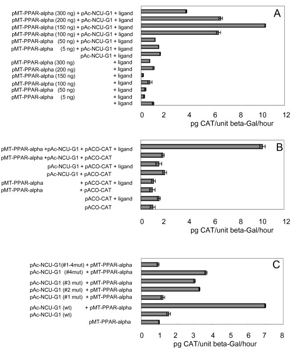
Results: NCU-G1 was found to be a highly conserved nuclear protein rich in proline with a molecular weight of approximately 44 kDa. It is localized on chromosome 1 and consists of 6 exons. Analysis of the amino acid sequence revealed no known transcription activation domains or DNA binding regions, however, four nuclear receptor boxes (LXXLL), and four SH3-interaction motives in addition to numerous potential phosphorylation sites were found. Two nuclear export signals were identified, but no nuclear localization signal. In man, NCU-G1 was found to be widely expressed at the mRNA level with especially high levels detected in prostate, liver and kidney. Electrophoretic mobility shift analysis showed specific binding of NCU-G1 to an oligonucleotide representing the footprint 1 element of the human cellular retinol-binding protein 1 gene promoter. NCU-G1 was found to activate transcription from this promoter and required presence of the footprint 1 element. In transiently transfected Drosophila Schneider S2 cells, we demonstrated that NCU-G1 functions as a co-activator for ligand-activated PPAR-alpha, resulting in an increased expression of a CAT reporter gene under control of the peroxisome proliferator-activated receptor-alpha responsive acyl-CoA oxidase promoter.

Conclusion: We propose that NCU-G1 is a dual-function protein capable of functioning as a transcription factor as well as a nuclear receptor co-activator.


Figures

Figure 1
Figure 1
Analysis of the NCU-G1 amino acid sequence. Panel A: Comparison of deduced NCU-G1 amino acid sequences from various species. Totally conserved amino acids are shown against a black background. Highly conserved amino acids are shown against a grey background. The positions of conserved prolines are marked with black bullets. Panel B: Potential functional motifs in human NCU-G1.
Figure 2
Figure 2
Endogenous NCU-G1 is a nuclear protein. Panel A: S2 cells were transfected with expression vectors for hNCU-G1 (pAc-NCU-G1) or its truncated version pAc-hNCU-G1(Δ exon 6). Whole cell extracts (20 μg/lane) were analyzed by Western blotting using preimmune serum (lanes 1 to 3) or an antiserum raised against a C-terminal 15 amino acid peptide (lanes 5 to 7). Lane 1 and 5: control, untreated S2 cells, lane 2 and 6: S2 cells expressing hNCU-G1, lane 3 and 7: S2 cells expressing hNCU-G1(Δ exon6). Panel B: Cytoplasmic and nuclear extracts (20 μg/lane) from JEG3, RPE and 293 cells were size-fractionated on 10% SDS-PAGE gels and analyzed by Western blotting using NCU-G1 antiserum. Lane 1: JEG3 cytosol, lane 2: JEG3 nuclear extract, lane 3: RPE cytosol, lane 4: RPE nuclear extract, lane 5: 293 cytosol, lane 6: 293 nuclear extract. Panel C: The membranes used in panel B were stripped and analyzed with an antibody against Sp1.
Figure 3
Figure 3
Human NCU-G1 binds to the FP1-element of the CRBP1 promoter. NCU-G1 was expressed as a fusion protein with GST in E. coli and purified. Interaction between GST-NCU-G1 and radiolabeled FP1-oligonucleotide was analysed by EMSA. Lane 1 shows 32P-labeled FP1 oligonucleotide (control). Lane 2 shows complex formation between GST-NCU-G1 and 32P-labeled FP1 oligonucleotide. Lane 3, 4, 5 and 6 show complex formation between GST-NCU-G1 and 32P-labeled FP1 oligonucleotide in the presence of 50-fold molar excess of unlabeled FP1 oligonucleotide, Ap1 oligonucleotide, Sp1 oligonucleotide or NF1 oligonucleotide, respectively. Ns = non-specific.
Figure 4
Figure 4
Human NCU-G1 activates transcription from the hCRBP1 promoter. Panel A: S2 cells were co-transfected with 250 ng pAc5.1/V5-His-LacZ, 350 ng phCRBP1-CAT and increasing amounts (2.5 ng – 50 ng) of pAc-hNCU-G1 as shown in the figure. Panel B: S2 cells were co-transfected with 250 ng pAc5.1/V5-His-LacZ, 350 ng plasmid expressing CAT under the control of various fragments of the hCRBP1 promoter as shown in the figure and 10 ng pAc-hNCU-G1. Panel C: S2 cells were co-transfected with 250 ng pAc5.1/V5-His-LacZ, 350 ng phCRBP1-CAT and 10 ng pAc-hNCU-G1 either wild-type or carrying mutated NR-boxes as shown in the figure. Panel D: S2 cells were cotransfected with 250 ng pAc5.1/V5-His-LacZ, 350 ng phCRBP1-CAT and 10 ng pAc-hNCU-G1 either wild-type or carrying various deletions as shown in the figure. Controls show expression of CAT in the absence of any NCU-G1. The cells were harvested 48 hrs after transfection. CAT was assayed by ELISA and β-galactosidase by enzyme activity. The data represent relative average values (pg CAT/unit β-galactosidase/hr +/- SE) from three independent experiments carried out in triplicates.
Figure 5
Figure 5
Human NCU-G1 functions as a ligand dependent and NR-box dependent co-activator for PPAR-alpha in S2 cells. Panel A: S2 cells were transiently transfected with 250 ng pAc5.1V5/His-LacZ, 350 ng of pACO-CAT, and increasing amounts of pMT-hPPARalpha, in the presence or absence of 10 ng of pAc-hNCU-G1. Twelve hrs after transfection WY14643 was added to 20 μM. The cells were harvested 56 hrs after transfection. CAT was assayed by ELISA and β-galactosidase by enzyme activity. The data represent relative average values (pg CAT/unit β-galactosidase/hr +/- SE) from three independent experiments carried out in triplicates. Panel B: S2 cells were transiently transfected with 250 ng pAc5.1V5/His-LacZ, 350 ng of pACO-CAT, together with pAc-hNCU-G1, pMT-hPPAR-alpha and WY14643 as indicated. The cells were treated and presented as in panel A. Panel C: S2 cells were transiently transfected with 250 ng pAc5.1V5/His-LacZ, 350 ng of pACO-CAT, 150 ng pMT-hPPAR-alpha as indicated and 10 ng of pAc-hNCU-G1, either wild-type or mutated as indicated in the figure. All cells were treated with 20 μM WY14643. The cells were harvested and data presented as in panel A.
Figure 6
Figure 6
Human NCU-G1 co-localises with PPAR-alpha in the nucleus. HeLa cells were transiently transfected with 0.8 μg pNCU-G1-EGFP and 0.8 μg pRFP-PPAR-alpha. Twenty hours after transfection WY14643 was added to 2 μM. Six hours later the live cells were analysed by confocal fluorescence microscopy. Panel A: Green fluorescence showing subcellular localisation of NCU-G1-EGFP. Panel B: Red fluorescence showing subcellular localisation of RFP-PPAR-alpha. Panel C: Merged picture. Arrows show foci of co-localisation.

References

    1. Mangelsdorf DJ. Vitamin A receptors. Nutr Rev. 1994;52:S32–44. - PubMed
    1. Tsai MJ, O'Malley BW. Molecular mechanisms of action of steroid/thyroid receptor superfamily members. Annu Rev Biochem. 1994;63:451–486. doi: 10.1146/annurev.bi.63.070194.002315. - DOI - PubMed
    1. Altucci L, Gronemeyer H. The promise of retinoids to fight against cancer. Nat Rev Cancer. 2001;1:181–193. doi: 10.1038/35106036. - DOI - PubMed
    1. Farias EF, Marzan C, Mira-y-Lopez R. Cellular retinol-binding protein-I inhibits PI3K/Akt signaling through a retinoic acid receptor-dependent mechanism that regulates p85-p110 heterodimerization. Oncogene. 2005;24:1598–1606. doi: 10.1038/sj.onc.1208347. - DOI - PubMed
    1. Cvetkovic D, Williams SJ, Hamilton TC. Loss of cellular retinol-binding protein 1 gene expression in microdissected human ovarian cancer. Clin Cancer Res. 2003;9:1013–1020. - PubMed

Publication types

MeSH terms