. 2008 Jan 4;375(1):301-15.
doi: 10.1016/j.jmb.2007.10.060. Epub 2007 Nov 1.

Biochemical and structural characterization of a novel family of cystathionine beta-synthase domain proteins fused to a Zn ribbon-like domain

Affiliations

Biochemical and structural characterization of a novel family of cystathionine beta-synthase domain proteins fused to a Zn ribbon-like domain

Michael Proudfoot et al. J Mol Biol. .

Abstract

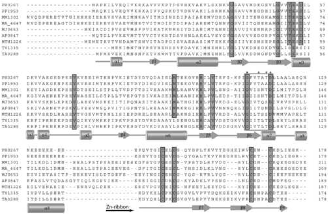
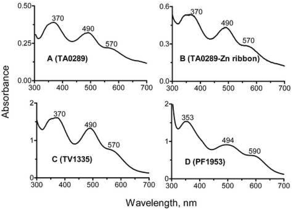
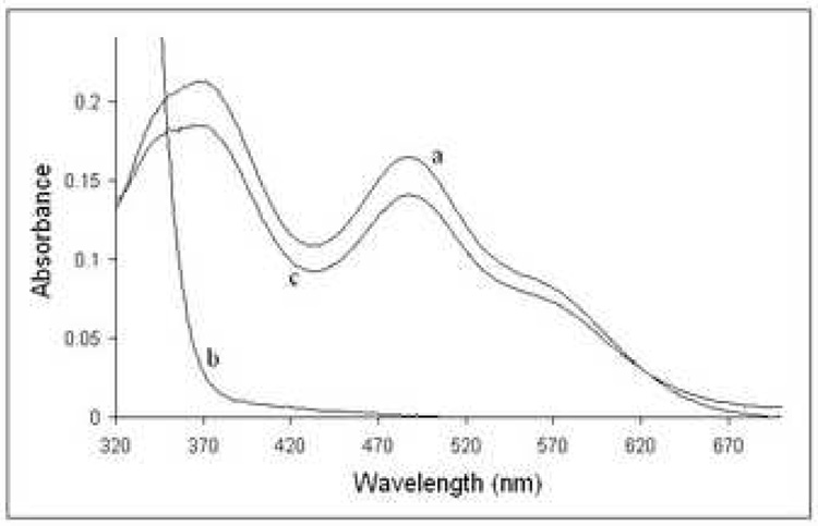
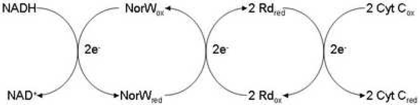
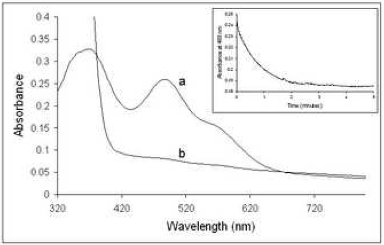
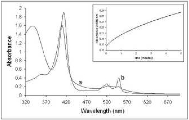
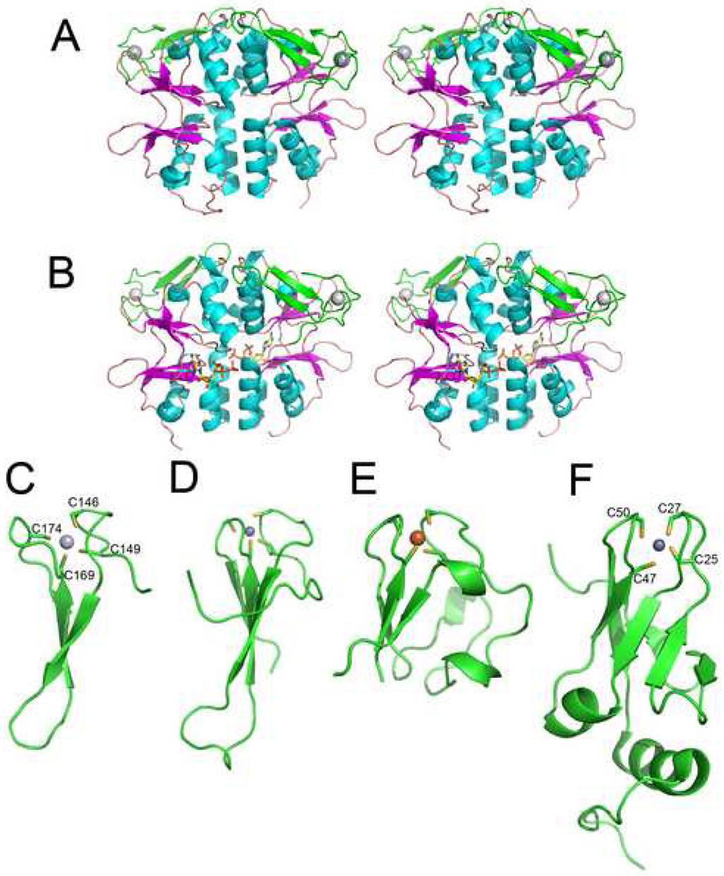
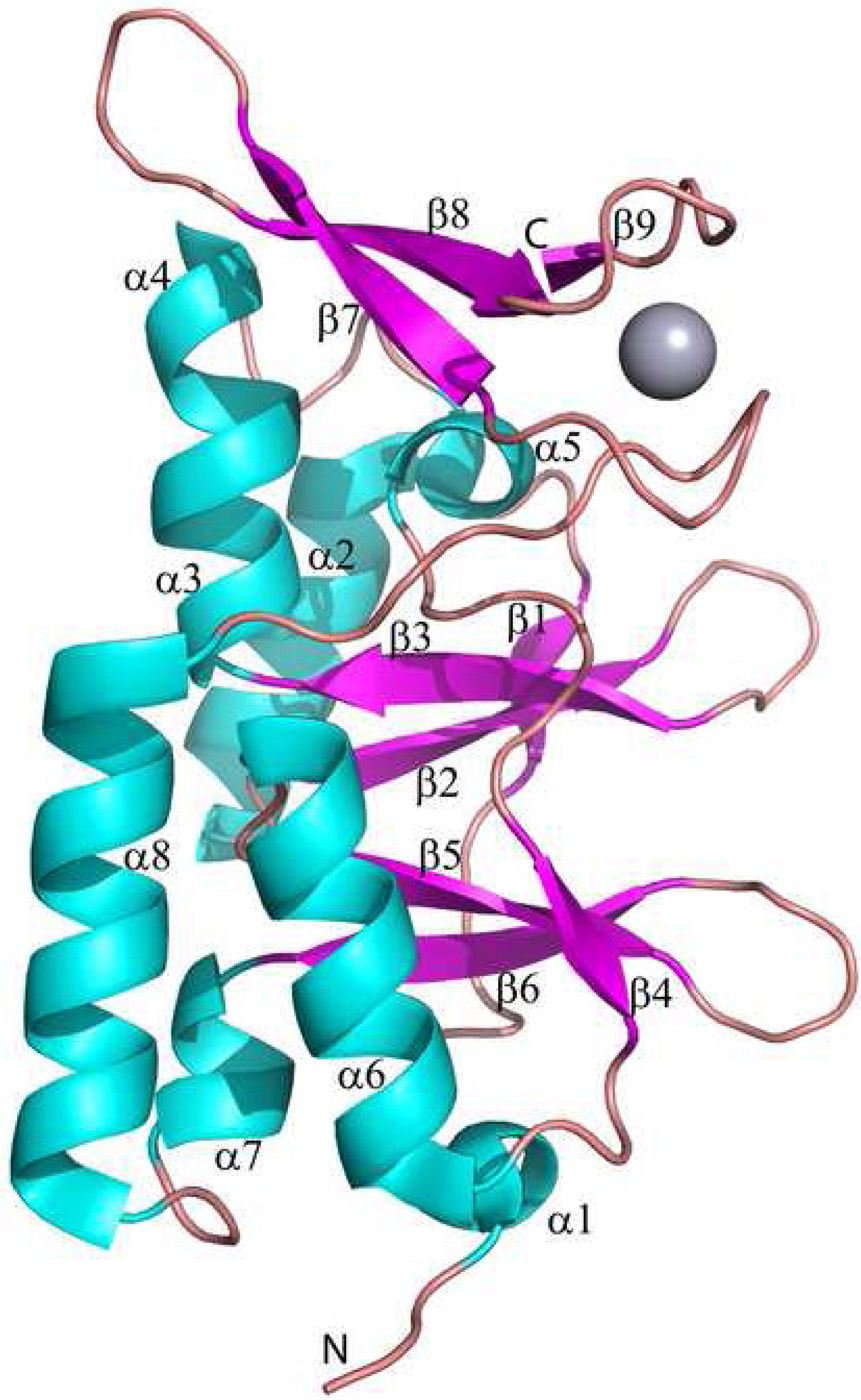
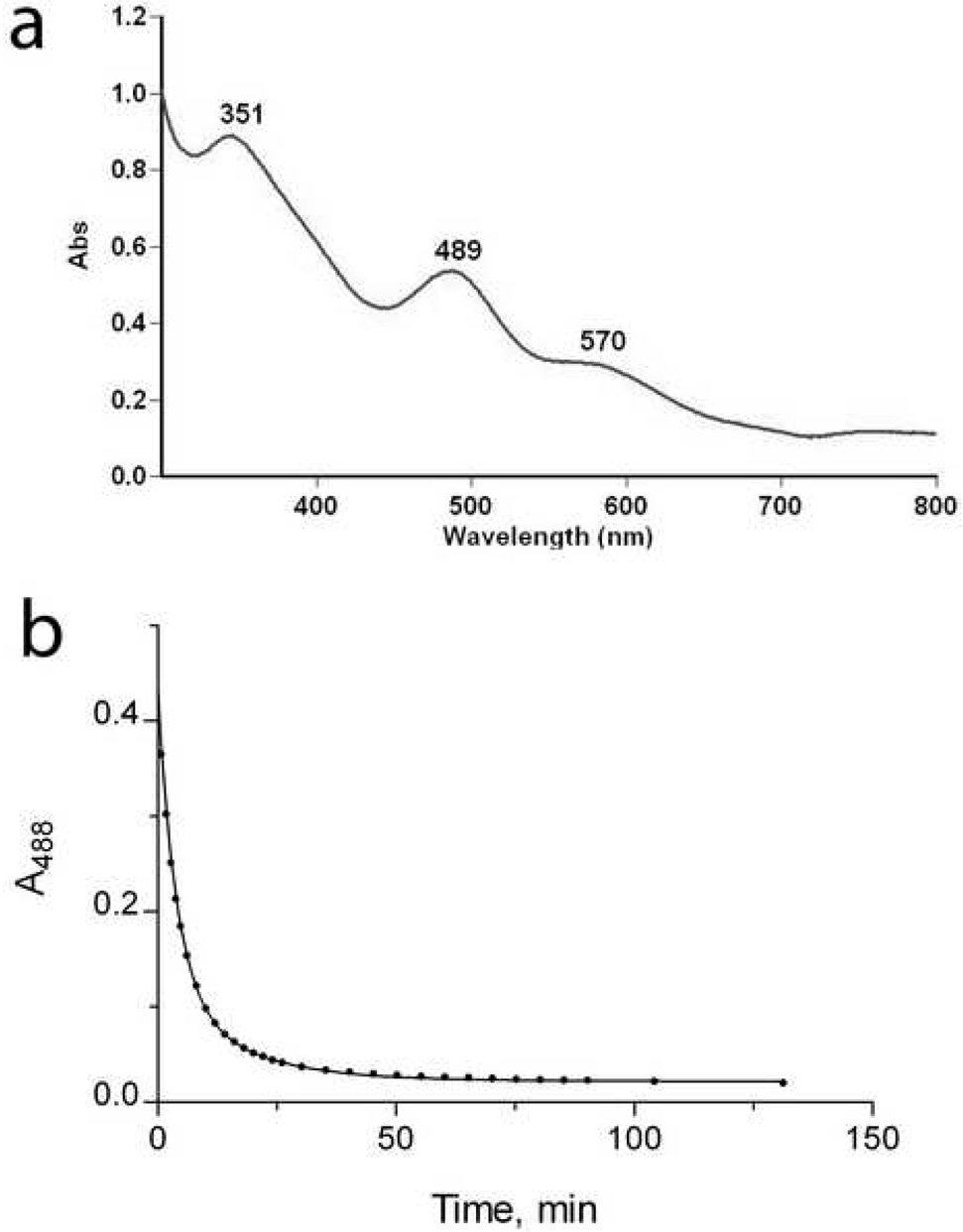
We have identified a novel family of proteins, in which the N-terminal cystathionine beta-synthase (CBS) domain is fused to the C-terminal Zn ribbon domain. Four proteins were overexpressed in Escherichia coli and purified: TA0289 from Thermoplasma acidophilum, TV1335 from Thermoplasma volcanium, PF1953 from Pyrococcus furiosus, and PH0267 from Pyrococcus horikoshii. The purified proteins had a red/purple color in solution and an absorption spectrum typical of rubredoxins (Rds). Metal analysis of purified proteins revealed the presence of several metals, with iron and zinc being the most abundant metals (2-67% of iron and 12-74% of zinc). Crystal structures of both mercury- and iron-bound TA0289 (1.5-2.0 A resolution) revealed a dimeric protein whose intersubunit contacts are formed exclusively by the alpha-helices of two cystathionine beta-synthase subdomains, whereas the C-terminal domain has a classical Zn ribbon planar architecture. All proteins were reversibly reduced by chemical reductants (ascorbate or dithionite) or by the general Rd reductase NorW from E. coli in the presence of NADH. Reduced TA0289 was found to be capable of transferring electrons to cytochrome C from horse heart. Likewise, the purified Zn ribbon protein KTI11 from Saccharomyces cerevisiae had a purple color in solution and an Rd-like absorption spectrum, contained both iron and zinc, and was reduced by the Rd reductase NorW from E. coli. Thus, recombinant Zn ribbon domains from archaea and yeast demonstrate an Rd-like electron carrier activity in vitro. We suggest that, in vivo, some Zn ribbon domains might also bind iron and therefore possess an electron carrier activity, adding another physiological role to this large family of important proteins.


Figures

Figure 1
Figure 1
Multiple sequence alignment of CBS-Zn ribbon-like proteins from various organisms. Highly conserved residues are highlighted in gray. The secondary structure elements derived from the structure of TA0289 are shown below the sequence alignment. The beginning of the Zn ribbon-like domain is indicated by a horizontal black arrow below the alignment. The residues marked with asterisks (and boxed) represent the predicted ribose-phosphate binding site.
Figure 2
Figure 2
Absorption spectra of purified CBS-Zn ribbon-like proteins. (A), TA0289; (B), Zn ribbon domain of TA0289; (C), TV1335; (D), PF1953.
Figure 3
Figure 3
Reduction of TA0289 by dithionite and reoxidation by air. 19 µM TA0289 in 50 mM Hepes-Na+ (pH 7.5), 0.2 M NaCl, and 10% glycerol (trace a) was scanned. A 50 mM stock of anaerobic sodium dithionite was then diluted 100-fold into the solution (to a final concentration of 500 µM), resulting in rapid reduction of TA0289 (trace b). After standing for several hours in air, a reoxidized spectrum (trace c) was obtained.
Figure 4
Figure 4
In vitro electron carrier assay for Rds and Zn ribbon-like proteins: scheme showing the electron transfer from NADH to cytochrome C via the E. coli NorW and Rd (or other electron carrier).
Figure 5
Figure 5
Anaerobic reduction of TA0289 by the E. coli NorW in the presence of NADH. A 24 µM anaerobic sample of TA0289 in a solution containing 50 mM Hepes-Na+ (pH 7.5), 0.2 M NaCl, 10% glycerol and 1 µM NorW was scanned (trace a). Reduction of TA0289 was achieved by the anaerobic addition of 200 µM NADH. The fully reduced spectrum is trace b. INSET: An absorbance trace at 488 nm following the time course for the reduction of 19 µM TA0289 in the presence of 200 µM NADH and 1 µM NorW in the same buffer, obtained from a single wavelength stopped flow experiment. The dotted line represents a fitted first order curve with a rate constant of 1.3 min−1.
Figure 6
Figure 6
Reduction of cytochrome C by TA0289 in the presence of NorW and NADH. A 200 mg ml−1 sample of cytochrome C in 50 mM Hepes-Na (pH 7.5), 0.2 M NaCl, and 10% glycerol in the presence of 1 µM NorW and 1.9 µM TA0289 was scanned (trace a). Reduction of cytochrome C was achieved by the anaerobic addition of 200 µM NADH. The fully reduced spectrum is trace b. INSET: An absorbance trace at 550 nm following the time course for the reduction of 200 µg ml−1 cytochrome C in the presence of 200 µM NADH, 1 µM NorW, and 1.9 µM TA0289 in the same buffer, obtained from a single wavelength stopped flow experiment.
Figure 7
Figure 7
Structures of TA0289 and other Zn ribbon domain proteins. (A), overall structure of the TA0289 dimer (stereo view). CBS domains are shown in orange (β-strands) and cyan (α-helices), Zn ribbon domains are shown in green, and metal atoms are shown as grey spheres. (B), stereo view of the TA0289 dimer shown with docked ATP from in silico analysis. (C), structure of the TA0289 Zn ribbon-like domain showing the tetrahedral coordination of the metal atom (Hg2+) by conserved cysteines (labeled). (D), structure of the Zn ribbon domain of the human transcriptional elongation factor TFIIS (1tfi). (E), structure of the P. furiosus rubredoxin PF1282 (1brf). (F), structure of KTI11 (1yop, 1yws). Metals are shown as: dark gray spheres (zinc; D and F), light gray sphere (mercury; C), and orange sphere (iron; E).
Figure 8
Figure 8
Structure of the TA0289 subunit. The secondary structure elements are numbered and shown in different colors (α-helices – cyan, β-strands – magenta, and loops – salmon). The position of the bound metal (Hg2+) is shown as a gray sphere.
Figure 9
Figure 9
Biochemical characterization of KTI11. (a), absorption spectrum of KTI11 purified from iron-supplemented cells. (b), anaerobic reduction of KTI11 (180 µM) by the E. coli NorW (20 nM) in the presence of NADH (0.2 mM). An absorbance trace at 488 nm following the time course for the reduction KTI11.

References

    1. Bateman A. The structure of a domain common to archaebacteria and the homocystinuria disease protein. Trends Biochem Sci. 1997;22:12–13. - PubMed
    1. Ignoul S, Eggermont J. CBS domains: structure, function, and pathology in human proteins. Am J Physiol Cell Physiol. 2005;289:C1369–C1378. - PubMed
    1. Scott JW, Hawley SA, Green KA, Anis M, Stewart G, Scullion GA, Norman DG, Hardie DG. CBS domains form energy-sensing modules whose binding of adenosine ligands is disrupted by disease mutations. J Clin Invest. 2004;113:274–284. - PMC - PubMed
    1. Bennetts B, Rychkov GY, Ng HL, Morton CJ, Stapleton D, Parker MW, Cromer BA. Cytoplasmic ATP-sensing domains regulate gating of skeletal muscle ClC-1 chloride channels. J Biol Chem. 2005;280:32452–32458. - PubMed
    1. Biemans-Oldehinkel E, Mahmood NA, Poolman B. A sensor for intracellular ionic strength. Proc Natl Acad Sci U S A. 2006;103:10624–10629. - PMC - PubMed

Publication types

MeSH terms