. 2008 Jan 15;314(2):297-308.
doi: 10.1016/j.yexcr.2007.10.010. Epub 2007 Oct 23.

Modulation of osteoblast gap junction connectivity by serum, TNFalpha, and TRAIL

Affiliations

Modulation of osteoblast gap junction connectivity by serum, TNFalpha, and TRAIL

Allison C Sharrow et al. Exp Cell Res. .

Abstract

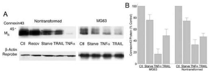
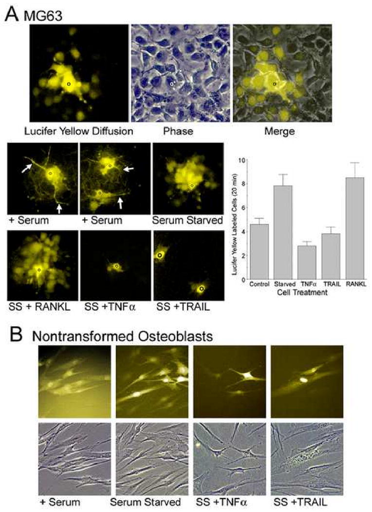
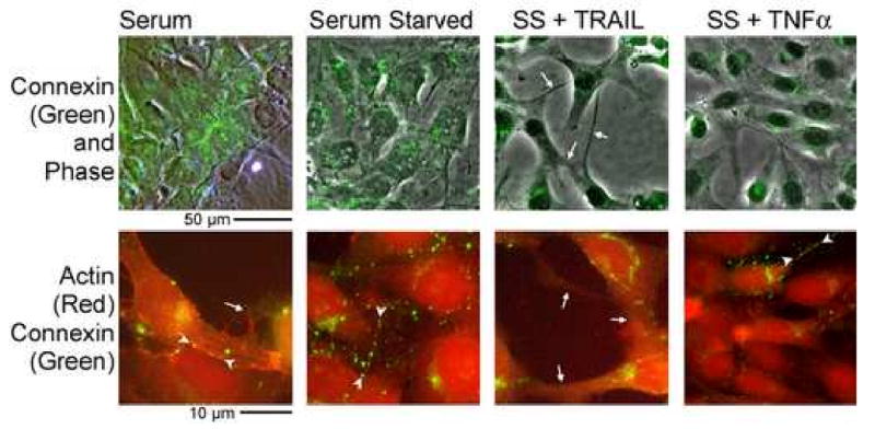
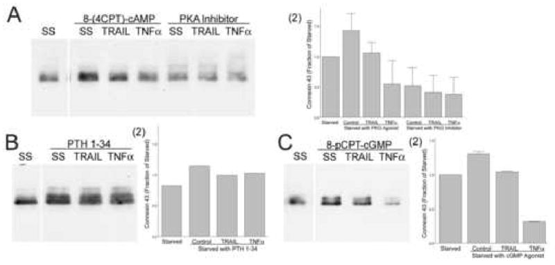
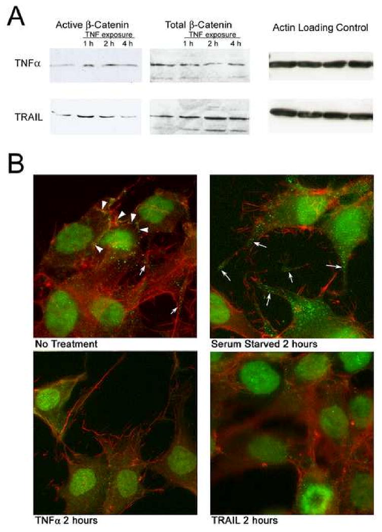
We studied the effects of serum growth factors and of TNF family proteins on osteoblast gap junction connectivity. Serum starvation of human MG63 osteosarcoma cells or nontransformed osteoblasts decreased connexin43 protein. TNFalpha or TRAIL reduced connexin43 further. Serum starvation redistributed gap junctions but did not reduce intercellular diffusion. In contrast, TNFalpha or TRAIL reduced gap junctions on cell processes and decreased intercellular diffusion. Effects of TNFs on connexin43 were mediated by lysosomal proteolysis. Activating analogs of cAMP increased connexin43 protein, but did not block effects of serum starvation, TNFalpha, or TRAIL on connexin43 protein. Connexin43 and connectivity recovered overnight if stimuli were withdrawn. Surprisingly, connexin43 mRNA increased in serum starvation and with TNFalpha or TRAIL. Since beta-catenin is a binding partner of connexin43, when connexin43 is degraded, beta-catenin activation may contribute to a reflexive increase in connexin43 transcription. We conclude that osteoblast connectivity is regulated by a multifactorial system that maintains intercellular connections. Serum starvation, TNFalpha and TRAIL augmented connexin43 degradation and connexin43 transcription. Cell-cell communication was maintained in serum starvation, which may model response to acute injury, but was sensitive to TNFs. These inflammatory agents mediated selective, reversible removal of connexin43 from cell processes.


Figures

Figure 1
Figure 1. Effect of TNFα and TRAIL on osteoblast survival and cell shape
A. Fas causes apoptosis in starved MG63 cells, but TRAIL and TNFα do not. Cells are shown after being serum starved 5 hours, treated with anti-Fas, TRAIL or TNFα and labeled with annexin V (green label) two hours later. Sporadic dead cells occurred with starvation (top left), while crosslinking with anti-human Fas (0.1 μg/ml, IgM) caused nearly quantitative apoptosis (top right). Apoptosis in 100 ng/ml TRAIL and TNFα (bottom left and right) was not significantly above background. Results at times up to 24 hours also showed no apoptosis above background with TRAIL and TNFα. Propidium iodide nuclear labeling occurred after annexin V labeling of Fas-crosslinked cells indicating apoptosis. Most annexin V labeled cells in other groups also labeled with propidium iodide. Photograph fields are 220 μm square. Apoptosis in Fas antibody was essentially quantitative, while cell death under other conditions was typically less than 5% and was not statistically different between serum starvation, TRAIL, and TNFα treatment. B. Effect of serum starvation and TRAIL on MG63 cell connectivity and comparison with nontransformed osteoblasts. The top left panel shows MG63 cells with epithelioid appearance when growing in RPMI 1640 with 10% fetal bovine serum. After five hours without serum (top right), the cell bodies retract leaving connecting processes between the cells. Addition of 10 ng/ml TRAIL reduces the connections (bottom left). Some cells have no connections and smaller cell diameter (arrows). Similar effects were seen in cells after addition of 10 ng/ml of TNFα (bottom right). Photograph fields are 100 μm square.
Figure 2
Figure 2. Connexin43 in osteoblasts is reduced by TRAIL or TNFα
A. Nontransformed osteoblasts (left) and MG63 cells (right) had reduced connexin43 protein after serum starvation and exposure to 10 ng/ml TRAIL or TNFα. The data are Western blots of cell lysates made after six hours of serum starvation followed by four hours of treatment, except for control cells which were not serum starved, serum starved controls (which had 10 hours without serum) and a culture which was serum starved and treated with TNFα but then allowed to recover in serum without added TNFα overnight (Recov, second lane of left panel). In some cases, the decrement in connexin43 was more than 90%, although not all experiments showed strong responses, and especially in TRAIL in long time-course experiments. This may reflect in part that inhibitors of TRAIL are secreted by osteoblasts (see Discussion). B. Response of connexin43 to serum starvation and TRAIL or TNFα. Five MG63 and five nontransformed osteoblast preparations were studied by Western blot as in (A). Optical density of connexin43 was measured and values normalized to control to allow comparisons to be displayed as mean ± SEM. Nontransformed and transformed cells were sensitive to TNFα and TRAIL, but effects of TRAIL were less consistent. Both MG63 and nontransformed cells make large quantities of TRAIL-binding soluble proteins [9]. The TNFα effect in both cells types relative to the starved cells is different at p < 0.01, while the TRAIL effect is variable and in this data set is different from serum starved in the MG63 cells at p < 0.05 and the difference in nontransformed cells has p ∼ 0.10. Data are from sub-confluent cells at 50-75% density.
Figure 3
Figure 3. Effect of TNFs on degradation of connexin43 and on connexin43 mRNA
A. Effect of proteasomal and lysosomal inhibitors. Western blots for Connexin43 in MG63 cells serum starved two hours and treated with 10 ng/ml of TRAIL or TNFα for four hours after the addition of 5 μM lactacystin, 1 mg/ml leupeptin, or 1 mM ammonium chloride. In lactacystin, which inhibits proteasomal degradation of ubiquitinated proteins, TRAIL and TNFα increased degradation of connexin43 essentially as in untreated cells (See Figure 2). Leupeptin and ammonia, however, eliminated almost all of the TNF-dependent connexin43 loss. Blots from three experiments with leupeptin or ammonium chloride gave similar results. Blots were reprobed for actin to show similar protein loads, and meaningful differences were not seen. Densitometry showed that decrements in connexin43 in lactacystin were not statistically different from levels shown in Fig 1B, while in in leupeptin or ammonium chloride, there was no statistically significant decrement in connexin43 with TRAIL or TNFα treatment. B. Effect of starvation and TNF-family proteins on connexin43, actin, and glycerol-3-phosphate dehydrogenase (GAPDH) mRNAs. Three independent real-time PCR reactions were run, each in duplicate or triplicate, measuring connexin43, two measuring actin and one measuring GAPDH. All gave similar results indicating that serum starvation increased connexin43 mRNA relative to α-actin and GAPDH. Within starved groups, the TNFs consistently further increased connexin43 mRNA over controls, but only 20-40% relative to serum starved individually with p values of ∼0.1 to ∼0.35. On the other hand, differences between controls and serum starvation relative to actin or GAPDH were both significant, p < 0.05. The bars show mean ± SEM, N=3, with controls normalized to 1.0.
Figure 4
Figure 4. Serum starvation, TNFα and TRAIL alter osteoblast connectivity
A. Dye diffusion in serum starved MG63 cells. Randomly selected individual cells in MG63 cells serum starved 4 hours were injected with 100 mM lucifer yellow (black circle). After 10 minutes, fluorescent images demonstrated intercellular diffusion. A typical result is shown in the top images with phase comparison. Approximately 20 cells labeled, most of them were adjacent but a few productive connections skip intermediate cells. Fields are 150 μm across. The lower panels show the effect of serum starvation and TNFs on dye diffusion. MG63 cells in growth media with serum (+ serum; three injections shown in two fields) gave diffusion involving ∼5 cells and showed prominent cell processes (arrows). In serum starved cultures intercellular processes were greatly narrowed, but several times as many cell bodies labeled. Both TRAIL or TNFα reduced dye diffusion but the TNF family protein RANKL, for which receptors are absent in osteoblasts, had no effect. 220 μm square fields. The graph shows results from a second experiment with numbers of cells labeled at 20 minutes after lucifer yellow injection in sequential cultures. The number of labeled cells was significantly less in serum starved cells exposed 2 hours to TNFα or TRAIL (p<0.05) than in serum starved or RANKL treated osteoblasts (n=5 except for RANKL, n=4). B. Dye diffusion in nontransformed osteoblasts. Nontransformed osteoblasts are less uniform than MG63 cells in shape. These cells had strong diffusion involving neighboring cells (left), even at low density. In these cells, serum starvation had smaller effects on cell shape. However, cell processes and connectivity were reduced by TNFα (third pair) and TRAIL (fourth pair). Each panel is 150 μm square.
Figure 5
Figure 5
Effect of TRAIL and TNFα on gap junctions and the actin cytoskeleton. MG63 cells were grown and labeled with phalloidin rhodamine for filamentous actin (red) and Alexafluor-488 anti-connexin43 (green) after three hour treatment with 10 ng/ml TNFα or TRAIL. Phase with connexin43 is shown at low power in the top frames and the bottom frames show actin and connexin43 at high power. Serum starvation reduced junctions between juxtaposed cell bodies and instead lines of connexons occurred on cell processes (arrowheads in the bottom frames of the serum and serum starved photographs). Connexin43 on cell processes was reduced in TRAIL or TNFα, with a large portion of the connexin43 labeling in a vacuolar distribution (masses of green material within cells). There was variation with occasional cells retaining processes with connexons, as shown by the arrowheads, in the bottom right frame, after TNFα treatment. The top panels are 150 μm across. The bottom panels are 40 μm across.
Figure 6
Figure 6
Effects of major kinase pathways and cytokines on osteoblast connexin43. All cultures were serum starved six hours prior to addition of stimuli, except that PTH was added at the time of serum starvation, with lysates collected after an additional two hours of incubation. TRAIL or TNFα were used at 10 ng/ml. Lysate proteins were measured and 25 μg loaded in each lane. A. Protein kinase A. Activating protein kinase A with the non-hydrolyzable cAMP analog 8-(4-CPT)-cAMP, 100 μM increased connexin43 relative to serum starvation, while PKA inhibitor peptide at 50 μM reduced connexin43 (left blots). The cAMP analogs did not abolish TRAIL or TNFα response. Similar PKA effects were seen in replicates. (2) Densitometry of the blot and a replicate, normalized to the density of serum starved cultures; Mean ± Range. B. PTH. PTH 1-34, 10 nM, increased connexin43 relative to serum starved cells, and blunted response of the TNFs (right lanes). (2) Densitometry of the blot normalized to the serum starved control. C. Protein kinase G. The nonhydrolyzable cGMP analog 8-pCPT-cGMP, 100 μM reduced connexin43. However, response to TNFα was not affected. Replicates gave the same result. (2) Densitometry of the blot and a replicate, normalized to the density of serum starved cultures; Mean ± Range.
Figure 7
Figure 7. Effect of TRAIL and TNFα on β- catenin activation in MG63 cells
A. Western blots of MG63 cultures after 2 hour serum starvation and subsequent treatment with 10 ng/ml of TNFα or TRAIL. An increase in active, serine37/threonine41 hypo-phosphorylated β-catenin was generally observed at 1-2 hours. β- catenin stimulates connexin43 transcription, in keeping with rapid recovery from TNF effects (Figure 2) and with increased connexin43 mRNA during TNF treatment. Also shown are total β- catenin and actin labeling controls. In some cases total β- catenin appeared to be decreased with treatment, but the connexin-associated β- catenin is a relatively small fraction of the total and this observation was not consistent. In some cases β-catenin activation appeared to be prolonged beyond 2 hours but this was also inconsistent. B. Effect of TRAIL and TNFα on peripheral distribution of β- catenin. In subconfluent MG63 cells, some β- catenin (green) is concentrated densely at cell borders (arrowheads), while occasional peripheral punctate concentrations are also seen. Under all conditions, there is also significant nuclear β- catenin. The cytoskeleton is also visualized using rhodamine phalloidin (red). After serum starvation, peripheral β- catenin is prominent on cell processes in patterns similar to those of connexin43, in keeping with the distribution of connexons in this manner and with β- catenin being a component of the connexon. Consistent with the degradation of most connexin43 in, the peripheral distribution of β- catenin was greatly reduced by10 ng/ml TRAIL or TNFα at 2 hours. Effects of the TNFs on β- catenin distribution in nontransformed cells are not illustrated but were were similar to those observed in MG63 cells. In these proliferating cell cultures, β- catenin is also seen in the nucleus due to unrelated activity in cell-cycle regulation; note the mitotic figure at the bottom of the TRAIL-treated culture, lower right panel (see text). Each frame is 70 μm square.

References

    1. Yellowley CE, Li Z, Zhou Z, Jacobs CR, Donahue HJ. Functional gap junctions between osteocytic and osteoblastic cells. J Bone Miner Res. 2000;15:209–217. - PubMed
    1. Cooper DM, Turinsky AL, Sensen CW, Hallgrimsson B. Quantitative 3D analysis of the canal network in cortical bone by micro-computed tomography. Anat Rec B New Anat. 2003;274:169–79. - PubMed
    1. Saez JC, Berthoud VM, Branes MC, Martinez AD, Beyer EC. Plasma membrane channels formed by connexins: their regulation and functions. Physiol Rev. 2003;83:1359–1400. - PubMed
    1. Eberhardt AW, Yeager-Jones A, Blair HC. Regional trabecular bone matrix degeneration and osteocyte death in femora of glucocorticoid treated rabbits. Endocrinology. 2001;142:1333–1340. - PubMed
    1. Furlan F, Lecanda F, Screen J, Civitelli R. Proliferation, differentiation and apoptosis in connexin43-null osteoblasts. Cell Commun Adhes. 2001;8:367–371. - PubMed

Publication types

MeSH terms