. 2007 Dec;47(27):3394-407.
doi: 10.1016/j.visres.2007.09.016.

Impaired photoreceptor protein transport and synaptic transmission in a mouse model of Bardet-Biedl syndrome

Affiliations

Impaired photoreceptor protein transport and synaptic transmission in a mouse model of Bardet-Biedl syndrome

Muhammad M Abd-El-Barr et al. Vision Res. 2007 Dec.

Abstract

Bardet-Biedl syndrome (BBS) is an oligogenic syndrome whose manifestations include retinal degeneration, renal abnormalities, obesity and polydactylia. Evidence suggests that the main etiopathophysiology of this syndrome is impaired intraflagellar transport (IFT). In this study, we study the Bbs4-null mouse and investigate photoreceptor structure and function after loss of this gene. We find that Bbs4-null mice have defects in the transport of phototransduction proteins from the inner segments to the outer segments, before signs of cell death. Additionally, we show defects in synaptic transmission from the photoreceptors to secondary neurons of the visual system, demonstrating multiple functions for BBS4 in photoreceptors.


Figures

Figure 1
Figure 1
Bbs4-null mice do not have photoreceptor ultrastructural defects at early age. A) Low power TEM vertical section of the retinas of a Bbs4 (+/+) mouse (AC) and Bbs4 (-/-) (B,D) at P20 (A,B) and 4 weeks of age (C,D). Note very similar dimensions of OS and nuclei numbers, although nuclei of the Bbs4 (-/-) are pyknotic Figures E-G are from a Bbs4 (+/+) mouse at 4 weeks of age E)Vertical TEM section illustrating connecting cilium, connecting outer segment (OS) and inner segment (IS) of a rod photoreceptor. F) Horizontal TEM section showing ‘9+0’ structure of connecting cilium. Note that microtubules are doublets. G) Horizontal TEM section through basal body showing similar ‘9+0’ structure. Note however that the 9 microtubules are triplets. Figures H-J are from a Bbs4(-/-) mouse at 4 weeks of age. H) Vertical TEM section illustrating connecting cilium, connecting outer segment (OS) and inner segment (IS) of a rod photoreceptor. I) Horizontal TEM section showing ‘9+0’ structure of connecting cilium. J) Horizontal TEM section through basal body showing similar ‘9+0’ structure. Scale bars, 25 μm (A,B,C,D), 1 μm (E), 250 nm (F), 100nm (G), 2 μm (H) 100 nm (I,J).
Figure 2
Figure 2
Immunolocalization of BBS4 reveals two distinct subcellular localizations. Immunofluorescence imaging of wild type (+/+) (8 weeks of age) frozen cryosections of the retina reveals that BBS4 (green) is located at both the inner segments and outer plexiform layers (OPL). Rhodopsin, the visual pigment of rod photoreceptors, which is known to be segregated to the outer segment (OS) is seen in red. B) BBS4 (green) shows similar localization as that of the plus-end motor protein kinesin II (red). Inset shows that these proteins do not colocalize in the OPL. C) Double labeling for BBS4 (green) and PKCα (red) shows that BBS4 is presynaptic to rod bipolar cells and does not colocalize. Scale bars 10 μm (A,B,C), 1 μm (inset, B), 5 μm (inset, C).
Figure 3
Figure 3
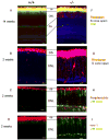
Immunofluorescent Imaging Reveals Selective Transport Defect in Photoreceptors of Bbs4-null mice. A,F) Immunofluorescence triple labeling of the retina shows proper localization of visual pigment, rhodopsin (red) and S cone opsin (blue) in a 14 week old wild type mouse (+/+) (A). Note that visual pigment is restricted to the outer segment (OS), which is where phototransduction occurs. The green stain, Peanut Agglutinin (PNA) binds to cone outer segment sheaths and pedicles. F) In a BBS4 (-/-) litter mate, there is evidence of considerable retinal degeneration as seen with the loss of nuclei in the outer nuclear layer (ONL). Of particular importance, note mislocalization of both rhodopsin (red) and S cone opsin (blue) in the ONL. B,G) Immunofluorescence double labeling for both rhodopsin and S cone opsin of reinas of (+/+) mouse (B) and (-/-) mouse (G) at 2 weeks of age. Note the mislocalization of both rhodopsin (red) and S cone opsin (blue) in the ONL., C,H) Immunofluorescence double labeling immunohistochemistry for L/M cone opsin (green) and peripherin/rds (red) of retinas of (+/+) mouse (C) and (-/-) mouse (h) at 2 weeks of age shows that both proteins are localized to the outer segments of photoreceptors of 2 week old Bbs4 (+/+) mice. Note in the Bbs4(-/-) mice (H), the mislocalization of the L/M opsin (green), but proper localization of peripherin/rds (red) D,I) Immunofluorescence double labeling for L/M cone opsin (green) and rom-1 (red) of retinas of (+/+) (D) and (-/-) (I) mousse at 2 weeks of age shows that both proteins are localized to the OS of photoreceptors of Bbs4 (+/+) mice, whereas in (-/-) mice, there is mislocalization of the L/M opsin (green), but proper localization of rom-1 (red) (I) E,J). Scale bar 10μm.
Figure 4
Figure 4
Impaired light-dependent translocations of transducin and arrestin in Bbs4-null mice. A) Segregation of rod transducin (green) and rod arrestin (red) in Bbs4 (+/+) in the dark. B) Bbs4 (+/+) mouse subjected to approximately 200 lux of light for 30 minutes. Note complete reciprocal translocation of transducin from the outer segment to the inner segment and arrestin from the inner segment to the outer segment. C) Note that segregation of transducin to the OS and arrestin to the IS is lost in Bbs4 (-/-) mouse also fixed in the dark. D) Bbs4(-/-) have incomplete translocation of both transducin (green) and arrestin (red) when subjected to the same duration and intensity of light as wild type mice (C). Note the perinuclear staining of the retained arrestin. Scale bar 10 μm. OS, Outer Segments; IS, Inner Segments; ONL, Outer Nuclear Layer; OPL, Outer Plexiform Layer.
Figure 5
Figure 5
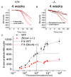
A) Fitting of ERG a-wave data at various intensities to Lamb-Pugh model of rod phototransduction for a 4 week old wild type mouse. The black traces are the portions of the raw data used in the fits, and the theoretical curves predicted by the fits are shown in red. B) Fitting of ERG a-wave data at various intensities to Lamb-Pugh model of rod phototransduction for a 4 week old Bbs4(-/-) mouse. The amplitudes of the a-waves have been normalized to that of the wild type so as to be more distinguishable. Although the amplitudes in (B) are greatly diminished, it is still quite possible to make good Lamb-Pugh fits to the data. At these intensities, kinetic parameters kA and t0 were indistinguishable between Bbs4(+/+) and Bbs4(-/-) mice (Table II). However, doing a similar fit to the highest intensity stimulus (approximately 8×10⁵ photoisomerizations/rod), there was a significant reduction in these parameters in the Bbs4(-/-) mice compared to the Bbs4(+/+) mice (Table II), likely due to decreased amount of photopigment (rhodopsin) and transducin (as shown by immunohistochemistry in Figure 3). C) Comparison of average b-wave amplitudes at various intensities for 4 week old wild type (n=7) and Bbs4(-/-) mice (n=6). In the first phase of intensities (< 100 photoisomerizations/rod) (labeled i), the b-waves can be fit with a Naka-Rushton fit (Table II). During the second phase (labeled ii), wild type (WT) mice exhibit growing b-waves, while Bbs4(-/-) mice do not.
Figure 6
Figure 6
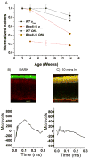
Discordance between Nuclei Numbers and ERG amplitudes can be partially explained by mislocalization of visual proteins. A) Comparison of nuclei numbers in ONL (circle symbols) compared to ERG amax (square symbols) with age reveals that amax decreases at a greater rate and at a greater extent than nuclei in the Bbs4(-/-) mice (red) as compared to wild type mice (black). B) Dark-adapted wild type retinas show clear segregation of transducin (green) to the OS and arrestin to the IS and ONL. At these conditions, the dark-adapted ERG amax is also of maximal amplitude (400-600μV). C) Subjecting wild type retinas to ambient light for 10 minutes causes gradual reciprocal translocation of arrestin to the OS and transducin to the IS. At these conditions, ERG amax amplitude are decreased (∼100-200 μV). Scale bars, 10 μm.
Figure 7
Figure 7
Western blot analysis shows that both the phototransduction proteins probed (rhodopsin, arrestin, transducin) and the structural protein peripherin/rds are full-length in the Bbs4-null mice at both P10 and P28. Moreover, there is a progressive decrease in the amounts of the phototransduction proteins with age. However, the amount of the structural protein peripherin/rds maintains the same with age, suggesting that the structural proteins, which we show are mislocalized (Figure 3) are degraded, but the structural proteins, such as peripherin/rds, which show proper localization (Figure 3) are not degraded.
Figure 8
Figure 8
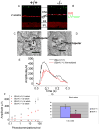
Immunofluroscence imaging of wild type (+/+) (A) and mutant (-/-) (B) retinas reveal correct localization of SV2 (red) to the OPL and IPL in both animals. Note the improper localization of L/M opsin (green) to the ONL and OPL in Bbs4(-/-) mice. C) Vertical TEM section showing typical synapse of rod to rod bipolar cell in a 4 week old Bbs4(+/+) mouse. Note presynaptic ribbon synapse, as well as post-synaptic triad consisting of one rod bipolar cell and two horizontal cells (hc). D) Vertical TEM section showing typical synapse of rod to rod bipolar cell in a 4 week old Bbs4(-/-) mouse. Note presynaptic ribbon synapse, as well as post-synaptic triad consisting of one rod bipolar cell and two horizontal cells (hc). E) Representative ERG of Bbs4(+/+) (black) and Bbs4(-/-) (red) shows that by normalizing the Bbs4(-/-) a-wave to that of the wild type, the b-wave is reduced in amplitude. F) ERG a-wave (circles) and b-wave (squares) for Bbs4(+/+) (black) and Bbs4(-/-) (red) mice at various intensities. G) Average b-to-a-wave ratio is significantly decreased in Bbs4(-/-) mice (p < 0.05, Student's t-test).
Figure 9
Figure 9
Proposed model of BBS4 function in photoreceptors. We have found evidence for two distinctive roles for BBS4 in the normal functioning of mammalian photoreceptors. The first involves the transport of phototransduction proteins such as rhodopsin and arrestin to the outer segment (OS). The second involves synaptic transmission at the axon terminal from photoreceptors to the secondary neurons of the visual pathway. Concerning the transport of proteins to the OS, our results suggest that there are at least two molecular mechanisms of transport. The first, of which the BBS4 protein plays an important role is in the transport of phototransduction proteins, such as the opsins and arrestin. The second mechanism involves structural proteins, such as peripherin/rds and rom-1, and is BBS4-independent

References

    1. Alfinito PD, Townes-Anderson E. Activation of mislocalized opsin kills rod cells: a novel mechanism for rod cell death in retinal disease. Proc Natl Acad Sci U S A. 2002 Apr 16;99(8):5655–60. - PMC - PubMed
    1. Ansley SJ, Badano JL, Blacque OE, Hill J, Hoskins BE, Leitch CC, Kim JC, Ross AJ, Eichers ER, Teslovich TM, Mah AK, Johnsen RC, Cavender JC, Lewis RA, Leroux MR, Beales PL, Katsanis N. Basal body dysfunction is a likely cause of pleiotropic Bardet-Biedl syndrome. Nature. 2003;425(6958):628–33. 9. - PubMed
    1. Baehr W, Karan S, Maeda T, Luo DG, Li S, Bronson JD, Watt CB, Yau KW, Frederick JM, Palczewski K. The function of guanylate cyclase 1 (GC1) and guanylate cyclase 2 (GC2) in rod and cone photoreceptors. J Biol Chem. 2007 Mar 23;282(12):8837–47. - PMC - PubMed
    1. Bascom RA, Manara S, Collins L, Molday RS, Kalnins VI, McInnes RR. Cloning of the cDNA for a novel photoreceptor membrane protein (rom-1) identifies a disk rim protein family implicated in human retinopathies. Neuron. 1992 Jun;8(6):1171–84. - PubMed
    1. Beales PL, Elcioglu N, Woolf AS, Parker D, Flinter FA. New criteria for improved diagnosis of Bardet-Biedl syndrome: results of a population survey. J Med Genet. 1999;36(6):437–46. - PMC - PubMed

Publication types

MeSH terms