Review
. 2008 Jan;29(1):114-27.
doi: 10.1016/j.yfrne.2007.08.005. Epub 2007 Oct 12.

Estrogen receptors: their roles in regulation of vasopressin release for maintenance of fluid and electrolyte homeostasis

Affiliations
Review

Estrogen receptors: their roles in regulation of vasopressin release for maintenance of fluid and electrolyte homeostasis

Celia D Sladek et al. Front Neuroendocrinol. 2008 Jan.

Abstract

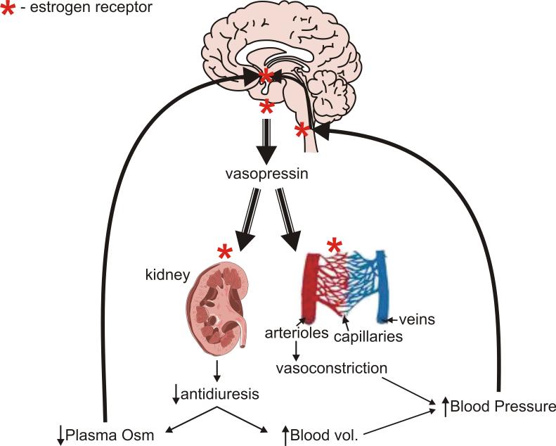
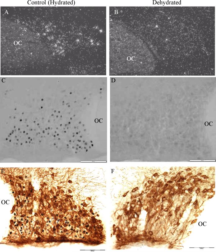
Long standing interest in the impact of gonadal steroid hormones on fluid and electrolyte balance has led to a body of literature filled with conflicting reports about gender differences, the effects of gonadectomy, hormone replacement, and reproductive cycles on plasma vasopressin (VP), VP secretion, and VP gene expression. This reflects the complexity of gonadal steroid hormone actions in the body resulting from multiple sites of action that impact fluid and electrolyte balance (e.g. VP target organs, afferent pathways regulating the VP neurons, and the VP secreting neurons themselves). It also reflects involvement of multiple types of estrogen receptors (ER) in these diverse sites including ERs that act as transcription factors regulating gene expression (i.e. the classic ERalpha as well as the more recently discovered ERbeta) and potentially G-protein coupled, membrane localized ERs that mediate rapid non-genomic actions of estrogen. Furthermore, altered expression of these receptors in physiologically diverse conditions of fluid and electrolyte balance contributes to the difficulty of using simplistic approaches such as gender comparisons, gonadectomy, and hormone replacement to assess the role of gonadal steroids in regulation of VP secretion for maintenance of fluid and electrolyte homeostasis. This review catalogs these inconsistencies and provides a frame work for understanding them by describing: (1) the effect of gonadal steroids on target organ responsiveness to VP; (2) the expression of multiple types of estrogen receptors in the VP neurons and in brain regions monitoring feedback signals from the periphery; and (3) the impact of dehydration and hyponatremia on expression of these receptors.


Figures

Figure 1
Figure 1
Sites of ER expression (either ERα or ERβ) in components of the system for maintaining fluid homeostasis. ER (*) is expressed in osmosensor regions of the anterior hypothalamus, the VP producing neurons of SON and PVN, the brainstem areas transmitting cardiovascular feedback information, and the VP target organs, the kidneys and arterioles. See text for discussion and references. See Table 2 for information on the type of ER at each location.
Figure 2
Figure 2
ERα expression in the osmosensitive components of the lamina terminalis. A. Diagram showing location of subfornical organ (SFO) and organum vasculosum of the lamina terminalis (OVLT), the two circumventricular organs in the anterior hypothalamus that monitor extracellular fluid osmolality. B.-E. Sections through SFO (B,C) and OVLT (D,E) from 48 hr water deprived rats were double stained for ERα (green) and Fos (red). Note the nuclear localization of both ERα and Fos that is indicative of their roles as transcription regulatory factors. Unlike some steroid receptors (e.g. glucocorticoid receptors), the nuclear localization of ERs is not dependent on a steroid ligand binding to the receptor, and evidence exists for ligand-independent, gene regulatory effects of ERs [50, 60]. The rectangles in B and D indicate regions shown at higher magnification in C and E respectively. Fos staining is indicative of neurons activated by the dehydration protocol, and numerous ERα positive neurons show Fos activation (yellow/orange, some indicted by white arrowheads in C and E). Scale bars, 50μm. Modified from [38, 76].
Figure 3
Figure 3
Effect of 3-β-diol, an androgenic metabolite of testosterone that acts as an ERβ agonist, on NMDA-stimulated VP release from explants of the hypothalamo-neurohypophyseal system (HNS). NMDA (50 μM) induced a significant, but transient increase in VP secretion (red triangle) that was blocked by inclusion of 3-β-diol (10nM) in the perifusate (green inverted triangle). [Two-way repeated measure ANOVA F=5.07, p=0.02; individual mean comparison at 5.3 hrs *p<0.05 versus time control (black circles) and NMDA alone]. Basal release (pg/ml was as follows: Time control, 142±23; NMDA, 163±17; NMDA+3-β-diol, 201±34.
Figure 4
Figure 4
ERα and Fos immunoreactivity in SFO of hydrated (sham-hydrated), 48 hr water deprived (sham-dehydrated), and hydrated or water deprived AV3V lesions rats (AV3V+hydrated and AV3V-dehydrated respectively) rats. Water deprivation induced a significant increase in Fos expression in SFO which was markedly exaggerated in the AV3V lesioned animals. Due to the impairment of osmotically stimulated VP secretion in these animals, they experience an extreme increase in plasma osmolality during water deprivation (380±10 vs 304±1 mOsm/kg H2O in sham-dehydrated). The density of ERα staining was greater in the periphery of SFO in the sham-dehydrated rats compared to hydrated rats (panel B; p<0.05), and both the density and number of ERα positive neurons increased in the AV3V+dehydrated rats with the increase in ERα positive neurons occurring in both the periphery and core of SFO (panel D; p<0.05). In spite of this dramatic increase in ERα expression, not all Fos positive cells (panel H) became ERα positive. Modified from [76].
Figure 5
Figure 5
ERα expression in SON in hydrated and dehydrated rats. A. and B. In situ hybridization for ERα mRNA reveals a decrease in ERβ mRNA following 72 hrs of 2% saline ingestion (see [78] for details). C. and D. ERβ immunohistochemistry in SON reveals a disappearance of ERβ protein in SON following 48 hrs of water deprivation. Note in the hydrated section that dense ERβ immunoreactivity is seen predominantly in the neurons positioned in the ventral portion of SON corresponding to the location of VP neurons. However, faint immunoreactivity is also present in neurons located more dorsally (e.g. in the region corresponding to OT neurons). E. and F. Double immunohistochemistry for OT (brown) and ERβ (black). Note the cytoplasmic localization of the OT immunoreactivity versus the nuclear localization of the ERβ immunoreactivity. In the hydrated example, dense ERβ immunoreactivity is not present in the OT neurons indicating it is primarily expressed in VP neurons in SON (see [78] for details and for pictures of VP/ERβ double immunostaining). Scale bar, 100 μm. OC, optic chiasm.

References

    1. Altura BM. Sex and estrogens and responsiveness of terminal arterioles to neurohypophyseal hormones and catecholamines. J Pharmacol Exp Ther. 1975;193(2):403–412. - PubMed
    1. Altura BM, Altura BT. Vascular smooth muscle and neurohypophyseal hormones. Federation Proceedings. 1977;36:1853–1860. - PubMed
    1. Berghorn KA, Knapp LT, Hoffman GE, Sherman TG. Induction of glucocorticoid receptor expression in hypothalamic magnocellular vasopressin neurons during chronic hypoosmolality. Endocrinology. 1995;136:804–811. - PubMed
    1. Brailoiu E, Dun SL, Brailoiu GC, Mizuo K, Sklar LA, Oprea TI, Prossnitz ER, Dun NJ. Distribution and characterization of estrogen receptor G protein-coupled receptor 30 in the rat central nervous system. J Endocrinol. 2007;193(2):311–321. - PubMed
    1. Burbach JPH, DeHoop MJ, Schmale H, Richter D, DeKloet ER, TenHaaf JA, DeWied D. Differential responses to osmotic stress of vasopressin-neurophysin mRNA in hypothalamic nuclei. Neuroendocrinology. 1984;39:582–584. - PubMed

Publication types