Review
. 2007 Dec 7;13(45):5971-8.
doi: 10.3748/wjg.v13.45.5971.

Endoscopic pancreatic duct stent placement for inflammatory pancreatic diseases

Review

Endoscopic pancreatic duct stent placement for inflammatory pancreatic diseases

Pier-Alberto Testoni. World J Gastroenterol. .

Abstract

The role of endoscopic therapy in the management of pancreatic diseases is continuously evolving; at present most pathological conditions of the pancreas are successfully treated by endoscopic retrograde cholangio-pancreatography (ERCP) or endoscopic ultrasound (EUS), or both. Endoscopic placement of stents has played and still plays a major role in the treatment of chronic pancreatitis, pseudocysts, pancreas divisum, main pancreatic duct injuries, pancreatic fistulae, complications of acute pancreatitis, recurrent idiopathic pancreatitis, and in the prevention of post-ERCP pancreatitis. These stents are currently routinely placed to reduce intraductal hypertension, bypass obstructing stones, restore lumen patency in cases with dominant, symptomatic strictures, seal main pancreatic duct disruption, drain pseudocysts or fluid collections, treat symptomatic major or minor papilla sphincter stenosis, and prevent procedure-induced acute pancreatitis. The present review aims at updating and discussing techniques, indications, and results of endoscopic pancreatic duct stent placement in acute and chronic inflammatory diseases of the pancreas.


Figures

Figure 1
Figure 1
Chronic obstructive pancreatitis involving the head of the gland. A: Pancreatography showing a distal stricture of the main pancreatic duct; B: Insertion of the guidewire into the main pancreatic duct; C: Placement of a 10F plastic stent over the guidewire.
Figure 2
Figure 2
Placement of a pancreatic stent after pancreatic sphincterotomy. A: Once biliary sphincterotomy has been performed leaving a guidewire into the common bile duct, pancreatic sphincter ablation is done with the standard sphincterotome; B: A plastic stent is then pushed over the guidewire into the main pancreatic duct; C: Pancreatic stent in place.
Figure 3
Figure 3
Chronic obstructive pancreatitis involving the head of the gland: insertion of an s-shaped stent into the main pancreatic duct.
Figure 4
Figure 4
Symptomatic, chronic obstructive pancreatitis at early stage with moderate dilation of the main pancreatic duct. A: Mechanical dilation of the pre-papillary stricture by a 10F dilator; B: Insertion of the guidewire; C: Insertion of a plastic 10F stent throughout the dilated stricture over the guidewire.
Figure 5
Figure 5
Chronic obstructive pancreatitis. A: Severe stricture of the main pancreatic duct with marked upstream dilation; B: Placement of a 10F plastic stent after mechanical dilation of the stricture.
Figure 6
Figure 6
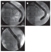
Refractory dominant stricture of the main pancreatic duct in chronic pancreatitis patient already treated by dilation and temporary stenting. Placement of two plastic stents. Endoscopic (A) and radiological (B) features.
Figure 7
Figure 7
Pancreas divisum with chronic obstructive pancreatitis at early stage. A: Extensive dorsal duct stricture with moderate upstream dilation of the pancreatic duct; B: Guidewire insertion into the dorsal duct throughout the minor papilla, without sphincterotomy; C: 7F plastic stent in place. Long-term stenting rather than minor papilla sphincterotomy appears an appropriate approach in this case with extensive dorsal duct stricture.
Figure 8
Figure 8
Chronic obstructive pancreatitis in incomplete pancreas divisum. A: Minor papilla stenosis associated with diffuse upstream dilation of the pancreatic duct and normal parietal morphology; B: Minor papilla sphincterotomy over-the-stent performed with a needle-knife sphincterotome. Minor papilla sphincterotomy rather than long-term stenting appears an appropriate approach in this case with stricture located at the level of the minor papilla.
Figure 9
Figure 9
Large pancreatic pseudocyst communicating with the main pancreatic duct: combined endoscopic trans-papillary and trans-gastric drainage. A: Contrast injection into the MPD shows a stricture of the main pancreatic duct at the level of the cyst; B: After pancreatic sphincterotomy, the stricture is dilated, a guidewire is inserted into the cyst cavity, and a plastic stent is placed; C and D: Under EUS guidance, a double pig-tail plastic stent is also placed by a single step procedure; D: X-ray imaging shows trans-papillary and trans-gastric stents at the end of the procedure.
Figure 10
Figure 10
Main pancreatic duct disruption with pancreatic juice leakage (A) successfully treated by plastic stent insertion bridging the leak (B).
Figure 11
Figure 11
Pancreatic duct leakage at the level of the tail of the gland, following surgical resection of neuroendocrine tumor. A: The leakage site is identified by contrast injection into the main pancreatic duct; B and C: Over a guidewire, a long plastic stent is inserted at the level of the tail of the gland.
Figure 12
Figure 12
Experimental winged 5 and 7F stents. The new stent design with a wing shape permits an adequate flow of pancreatic juice even alongside the stent and does not compress the duct over its entire circumference, thus avoiding the risk of impaired drainage of pancreatic juice and mechanical trauma to the duct.

References

    1. Ishihara T, Yamaguchi T, Seza K, Tadenuma H, Saisho H. Efficacy of s-type stents for the treatment of the main pancreatic duct stricture in patients with chronic pancreatitis. Scand J Gastroenterol. 2006;41:744–750. - PubMed
    1. Widdison AL, Alvarez C, Karanjia ND, Reber HA. Experimental evidence of beneficial effects of ductal decompression in chronic pancreatitis. Endoscopy. 1991;23:151–154. - PubMed
    1. Boerma D, van Gulik TM, Rauws EA, Obertop H, Gouma DJ. Outcome of pancreaticojejunostomy after previous endoscopic stenting in patients with chronic pancreatitis. Eur J Surg. 2002;168:223–228. - PubMed
    1. Adler DG, Lichtenstein D, Baron TH, Davila R, Egan JV, Gan SL, Qureshi WA, Rajan E, Shen B, Zuckerman MJ, et al. The role of endoscopy in patients with chronic pancreatitis. Gastrointest Endosc. 2006;63:933–937. - PubMed
    1. Díte P, Ruzicka M, Zboril V, Novotný I. A prospective, randomized trial comparing endoscopic and surgical therapy for chronic pancreatitis. Endoscopy. 2003;35:553–558. - PubMed