

Brain anatomy differences in childhood stuttering
- PMID: 18023366
- PMCID: PMC2731627
- DOI: 10.1016/j.neuroimage.2007.09.067
Brain anatomy differences in childhood stuttering
Abstract
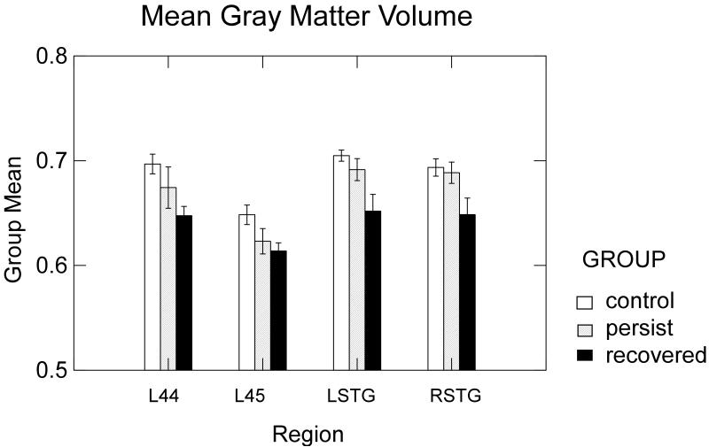
Stuttering is a developmental speech disorder that occurs in 5% of children with spontaneous remission in approximately 70% of cases. Previous imaging studies in adults with persistent stuttering found left white matter deficiencies and reversed right-left asymmetries compared to fluent controls. We hypothesized that similar differences might be present indicating brain development differences in children at risk of stuttering. Optimized voxel-based morphometry compared gray matter volume (GMV) and diffusion tensor imaging measured fractional anisotropy (FA) in white matter tracts in 3 groups: children with persistent stuttering, children recovered from stuttering, and fluent peers. Both the persistent stuttering and recovered groups had reduced GMV from normal in speech-relevant regions: the left inferior frontal gyrus and bilateral temporal regions. Reduced FA was found in the left white matter tracts underlying the motor regions for face and larynx in the persistent stuttering group. Contrary to previous findings in adults who stutter, no increases were found in the right hemisphere speech regions in stuttering or recovered children and no differences in right-left asymmetries. Instead, a risk for childhood stuttering was associated with deficiencies in left gray matter volume while reduced white matter integrity in the left hemisphere speech system was associated with persistent stuttering. Anatomical increases in right hemisphere structures previously found in adults who stutter may have resulted from a lifetime of stuttering. These findings point to the importance of considering the role of neuroplasticity during development when studying persistent forms of developmental disorders in adults.
Figures


References
-
- Ambrose NG, Yairi E. Normative disfluency data for early childhood stuttering. J Speech Lang Hear Res. 1999;42:895–909. - PubMed
-
- Amunts K, Weiss PH, Mohlberg H, Pieperhoff P, Eickhoff S, Gurd JM, Marshall JC, Shah NJ, Fink GR, Zilles K. Analysis of neural mechanisms underlying verbal fluency in cytoarchitectonically defined stereotaxic space--the roles of Brodmann areas 44 and 45. Neuroimage. 2004;22:42–56. - PubMed
-
- Ashburner J, Friston KJ. Voxel-based morphometry--the methods. Neuroimage. 2000;11:805–821. - PubMed
-
- Basser PJ, Pierpaoli C. Microstructural and Physiological Features of Tissues Elucidated by Quantitative-Diffusion-Tensor MRI. (Series B).Journal of Magnetic Resonance. 1996;111:209–219. - PubMed
Publication types
MeSH terms
Grants and funding
LinkOut - more resources
Full Text Sources
Medical
