Review
. 2007 Dec;17(6):633-40.
doi: 10.1016/j.sbi.2007.09.011. Epub 2007 Nov 19.

Intrinsic dynamics of enzymes in the unbound state and relation to allosteric regulation

Affiliations
Review

Intrinsic dynamics of enzymes in the unbound state and relation to allosteric regulation

Ivet Bahar et al. Curr Opin Struct Biol. 2007 Dec.

Abstract

In recent years, there has been a surge in the number of studies exploring the relationship between proteins' equilibrium dynamics and structural changes involved in function. An emerging concept, supported by both theory and experiments, is that under native state conditions proteins have an intrinsic ability to sample conformations that meet functional requirements. A typical example is the ability of enzymes to sample open and closed forms, irrespective of substrate, succeeded by the stabilization of one form (usually closed) upon substrate binding. This ability is structure-encoded, and plays a key role in facilitating allosteric regulation, which suggests complementing the sequence-encodes-structure paradigm of protein science by structure-encodes-dynamics-encodes-function. The emerging connection implies an evolutionary role in selecting/conserving structures based on their ability to achieve functional dynamics, and in turn, selecting sequences that fold into such 'apt' structures.


Figures
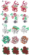
Figure 1
Figure 1. Experimental evidence for conformational diversity of folded proteins and comparison with theoretical predictions
(A) Three conformations of cyclin dependent kinases (CDKs) adopted in the free form (middle), and in the presence of two different substrates, an inhibitor (INK4; left) and its activator (cylin; right). The corresponding Protein Data Bank (PDB) codes are 1bi7, 1hcl and 1fin, in reading order. Colors refer to N-lobe (purple), C-lobe (red), hinge residues (orange) and activation loop (cyan). Both activation and inhibition involve conformational changes in and around the catalytic cleft. The activation loop (cyan) rotates towards the substrate (not shown). (B) Alternative conformations of HIV-1 RT. RT is composed of two subunits, p66 and p51 (wheat); the p66 subunit consists of two domains, polymerase and RNase H (blue); and the polymerase domain contains four subdomains, thumb (red), fingers (blue), palm (pink), connection (green). Comparison of the inhibitor-bound (nevirapine; space filling form in yellow; left), unliganded (middle) and DNA-bound (right) forms a (PDB codes 1rth, 1dlo and 2hmi) shows domain movements. (C) The left two diagrams display the free (left) and substrate-bound (middle) forms of adenylate kinase (AK) (respective PDB codes: 4ake and 1ake), and the diagram on the right is a reconfigured form of the free enzyme computed by deforming the unbound structure along the lowest frequency ANM mode (mode 1)[18]. AK contains three domains: core (white), lid (cyan), and AMP-binding (green) domains. The substrate is shown in orange, space-filling representation. In the substrate-bound form as well as the model on the right, the lid approaches the core. (D) Comparison of the T (tense, unliganded) (left) and R2 (relaxed, CO-bound) (middle) forms of hemoglobin (Hb) (respective PDB codes: 1a3n and 1bbb), and the model (right) calculated [19] by deforming the T form along ANM mode 2. The model approximates the experimentally observed torsion of the α2β2 dimer (front, colored) with respect to the α1β1 dimer (bottom, gray). (E) Comparison of the ribosome structure experimentally determined (middle), and two conformers (left and right) sampled by fluctuations along ANM mode 3 [62], approximating the ratchet-like rearrangement of the 70S subunit (green) with respect to the 30S (maroon), suggested by experimental data (see also the original work of [63]).
Figure 2
Figure 2. Combination of a pre-existing equilibrium and post-binding rearrangement
The protein (E) originally samples an ensemble of conformations, accessible via fluctuations in the global energy well (top panel), occasionally involving passages between substates within the well. The well is approximated by a harmonic potential (dashed curve) in coarse-grained NMA. Two conformations/substates are schematically shown, which are in a dynamic equilibrium prior to substrate (S) binding, as shown on the left box (bottom). The substrate (S) selects the conformer(s) that allows for optimal interaction (conformational selection). The stabilization of the final complex is subject to further rearrangement (induced fit) to stabilize the final bound form. The energy profile/landscape of the protein E in the presence of the substrate differs from its unbound form (top right diagram) inducing a shift in the population of the substates in favor of the conformer that binds the substrate.

References

    1. Hammes-Schiffer S, Benkovic SJ. Relating protein motion to catalysis. Annu Rev Biochem. 2006;75:519–541. This is an extensive review of experimental and theoretical evidence supporting the interplay between conformational dynamics of enzymes and their catalytic activities, with focus on two enzymes, dehydrofolate reductase and liver alcohol dehydrogenase. A network of coupled motions involving both fast thermal vibrations and low frequency modes is pointed out to modulate the interactions between enzyme, substrate and cofactor at the active site, facilitating the hydride transfer reaction. - PubMed
    1. Changeux JP, Edelstein SJ. Allosteric mechanisms of signal transduction. Science. 2005;308:1424–1428. This is an insightful review of the progress made in the last forty years in understanding the allosteric mechanisms of signal tranduction. The basic concepts underlying the MWC model are summarized, such as the pre-disposition of symmetric oligomeric structures to undergo cooperative changes and the occurrence of reversible transitions, or spontaneous ‘switches’ between discrete conformations, accessible in the absence of ligand. The importance of binding ligands at strategic locations such as interfaces between subunits or along symmetry axes is emphasized. - PubMed
    1. Bahar I, Rader AJ. Coarse-grained normal mode analysis in structural biology. Curr Opin Struct Biol. 2005;15:586–592. - PMC - PubMed
    1. Tama F, Brooks CL. Symmetry, form, and shape: guiding principles for robustness in macromolecular machines. Annu Rev Biophys Biomol Struct. 2006;35:115–133. An excellent review where the authors draw attention to the utility of multi-resolution methods based on elastic network models for understanding the functional dynamics of biological systems. They highlight the role of shape and form in the defining robust modes of motions. - PubMed
    1. Koshland DE. Application of a theory of enzyme specificity to protein synthesis. Proc Natl Acad Sci. 1958;44:98–104. - PMC - PubMed

LinkOut - more resources