. 2008 Mar 1;94(5):1600-12.
doi: 10.1529/biophysj.107.117580. Epub 2007 Nov 16.

Substrate binding and formation of an occluded state in the leucine transporter

Affiliations

Substrate binding and formation of an occluded state in the leucine transporter

Leyla Celik et al. Biophys J. .

Abstract

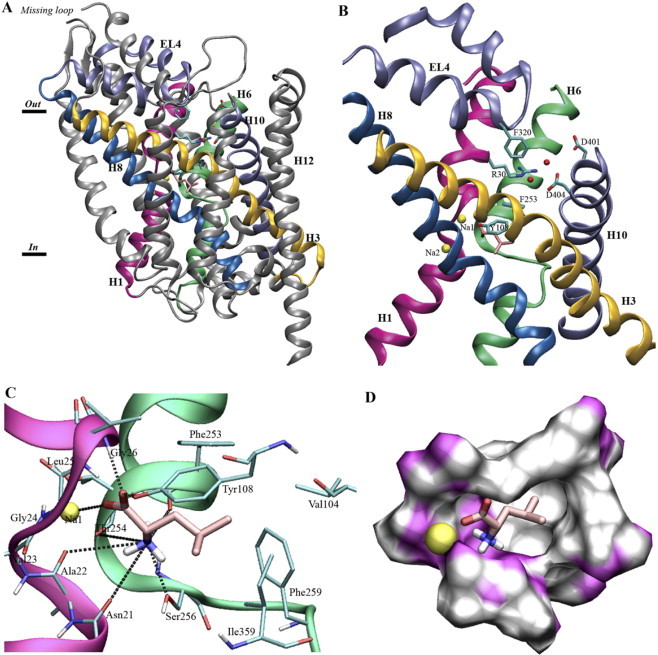
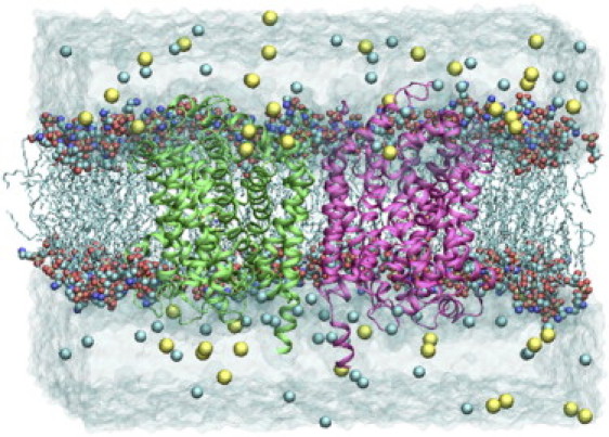
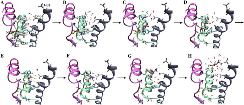
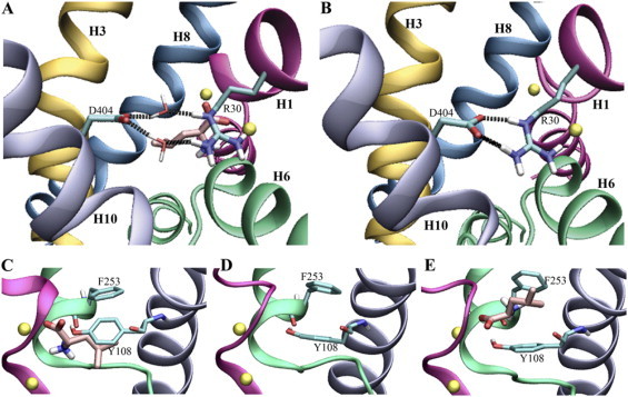
Translocation through the extracellular vestibule and binding of leucine in the leucine transporter (LeuT) have been studied with molecular dynamics simulations. More than 0.1 mus of all-atom molecular dynamics simulations have been performed on different combinations of LeuT, bound substrate, and bound structural Na(+) ions to describe molecular events involved in substrate binding and in the formation of the occluded state and to investigate the dynamics of this state. Three structural features are found to be directly involved in the initial steps of leucine transport: a Na(+) ion directly coordinated to leucine (Na-1), two aromatic residues closing the binding site toward the extracellular vestibule (Tyr-108 and Phe-253), and a salt bridge in the extracellular vestibule (Arg-30 and Asp-404). These features account for observed differences between simulations of LeuT with and without bound substrate and for a possible pathway for leucine binding and thereby formation of the occluded LeuT binding site.


Figures

Figure 1
Figure 1
(A) The crystal structure of LeuT. The 12 TM α-helices in LeuT are bundled together in a unique spiral-like fold. (B) The two unwound helices, H1 (magenta) and H6 (green), as well as helices H3 (yellow) and H8 (blue) are important for substrate binding and transport. H10 (light blue) and the extracellular loop 4 (EL4) (light blue) provide residues close to the suspected pathway. The Arg-30–Asp-404 salt bridge is water (red balls) mediated. (C) Substrate binding in LeuT. Snapshot of Leu (pink) and interacting residues (cyan) together with Na-1 (yellow). Note how residues interacting with the substrate functional groups are located in the unwound parts of H1 (magenta) and H6 (green). Possible hydrogen bonds to Leu(N) are marked with heteroatom distance. (D) The substrate, Leu (pink), and Na-1 (yellow sphere) in a surface representation of the LeuT binding site. The front of the figure is cut away to clearly show the hydrophobic (white) and hydrophilic (magenta) duality of the binding site.
Figure 2
Figure 2
The simulated system from a side view. The two transporter monomers are depicted in green and magenta cartoons, lipid tails in cyan lines, lipid heads in CPK, water in transparent, and ions as van der Waals spheres in yellow (Na+) and cyan (Cl). The limited interaction between the two monomers is through H11 and H12, which are not directly involved in substrate binding or transport, suggesting that the two monomers function independently.
Figure 3
Figure 3
The mechanism and steps involved in substrate (un-)binding. (A) Leu bound in the closed binding site. (B) Hydrogen bonds to Asn-21 and Ala-22 are released, and the binding site starts to open. (C) Hydrogen bonds to Ser-256 and Thr-254 are released, and the Leu side chain protrudes from the aromatic lid. (D) The ionic interaction to Na-1 is broken. (E) The hydrogen bond to Phe-253 is released. (F) Hydrogen bonds to Gly-26 and Tyr-108 are released, and the substrate is free of the binding site. Instead, interactions are found to the extracellular salt bridge. (G) Leu is turning around in the vestibule as a result of ionic interactions. (H) Leu is outside LeuT and solvated.
Figure 4
Figure 4
Water-mediated (A) and direct (B) interaction in the salt bridge between Arg-30 and Asp-404 in the extracellular vestibule. The aromatic lid on the extracellular side of the binding site can be either closed (C) or open (D), depending on the presence/absence of Leu. The open lid can also be seen during SMD simulation (E).

References

    1. Torres G.E., Gainetdinov R.R., Caron M.G. Plasma membrane monoamine transporters: structure, regulation and function. Nat. Rev. Neurosci. 2003;4:13–25. - PubMed
    1. Hahn M.K., Blakely R.D. Monoamine transporter gene structure and polymorphisms in relation to psychiatric and other complex disorders. Pharmacogenomics J. 2002;2:217–235. - PubMed
    1. Masson J., Sagne C., Hamon M., Mestikawy S.E. Neurotransmitter transporters in the central nervous system. Pharmacol. Rev. 1999;51:439–464. - PubMed
    1. Owens M.J., Nemeroff C.B. Role of serotonin in the pathophysiology of depression: focus on the serotonin transporter. Clin. Chem. 1994;40:288–295. - PubMed
    1. Rudnick G. Mechanisms of biogenic amine neurotransmitter transporters. In: Reith M.E.A., editor. Neurotransmitter transporters: structure, function, and regulation. 2nd edition. Humana Press; Totowa, NJ: 2002. pp. 25–52.

Publication types

MeSH terms

LinkOut - more resources