. 2007 Nov 27;104(48):18999-9004.
doi: 10.1073/pnas.0709387104. Epub 2007 Nov 16.

X-ray structure of EmrE supports dual topology model

Affiliations

X-ray structure of EmrE supports dual topology model

Yen-Ju Chen et al. Proc Natl Acad Sci U S A. .

Abstract

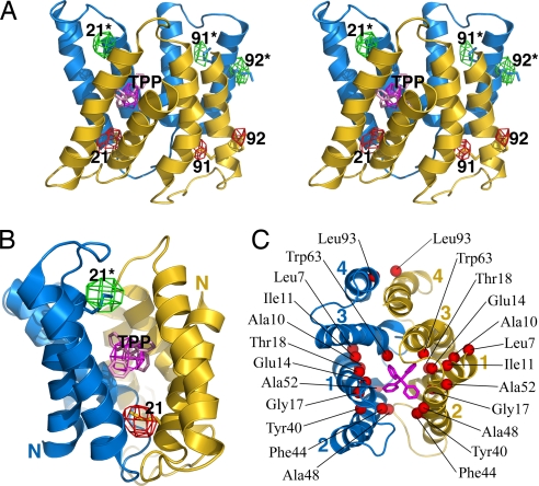
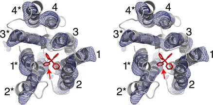
EmrE, a multidrug transporter from Escherichia coli, functions as a homodimer of a small four-transmembrane protein. The membrane insertion topology of the two monomers is controversial. Although the EmrE protein was reported to have a unique orientation in the membrane, models based on electron microscopy and now defunct x-ray structures, as well as recent biochemical studies, posit an antiparallel dimer. We have now reanalyzed our x-ray data on EmrE. The corrected structures in complex with a transport substrate are highly similar to the electron microscopy structure. The first three transmembrane helices from each monomer surround the substrate binding chamber, whereas the fourth helices participate only in dimer formation. Selenomethionine markers clearly indicate an antiparallel orientation for the monomers, supporting a "dual topology" model.


Conflict of interest statement

The authors declare no conflict of interest.

Figures

Fig. 1.
Fig. 1.
Biochemical characterization of EmrE expressed in vivo (filled circles) and in vitro (open circles). The data shown are representative experiments performed in NG; error bars show the standard deviation of three replicates done in parallel. (A) Binding of EmrE to TPP, as assayed by tryptophan fluorescence quenching. (Inset) Typical emission spectra of 25 μM EmrE at the indicated concentrations of added TPP. Plotting the percent quenching at 320 nm vs. the added TPP shows saturation curves in the main panel. The total concentration of TPP binding sites was estimated from the intersection of the two dashed lines, indicated by the arrow (≈12.5 μM). (B) Binding of EmrE to ethidium, measured by fluorescence anisotropy. The data were fit to a simple binding model (see Materials and Methods). (C) Competition assays measuring TPP binding to EmrE. Note the near-superposition of the curves for in vivo and in vitro-expressed EmrE in all three panels.
Fig. 2.
Fig. 2.
Structure determination of EmrE. (A) Experimental density for one apo EmrE monomer at 4.5-Å resolution. Anomalous Hg density (4σ), marking the positions of cysteine residues, is shown in red. The protein is shown in Cα trace and rendered in a color gradient, from green at the N terminus to yellow at the C terminus. (B) Ribbon representation of the distorted apo EmrE dimer. One monomer is rendered in color gradient with the helices labeled, and the other monomer is shown in gray. The approximate dimensions of a lipid bilayer are shown by the gray shading. (C) Views of the two apo EmrE monomers, with TM helices labeled. Note the extended configuration of the TM4 helices, which project away from the main body of the dimer. (D) Experimental density for one monomer of the EmrE-TPP complex at 3.8 Å (C2 crystal form), contoured at 1σ. Anomalous Se density (3σ) is shown in red. (E) Side view of the EmrE-TPP dimer (C2 form), with the dimensions of the lipid bilayer indicated. One monomer is colored in gradient and labeled, and the other is in gray. The bound TPP is colored red. Density for the colored monomer terminates at residue 105. (F) Views of the two monomers (P21 form), which are essentially the same as the C2 monomers, except for the shorter TM helices, which terminate at the indicated residues. Full-length EmrE has 110 amino acid residues. Note that the superhelical twists of TM1–3 are similar in the apo and TPP-bound forms but that the helix packing interactions and monomer–monomer interactions differ.
Fig. 3.
Fig. 3.
EmrE binds TPP as an antiparallel dimer. (A) Stereoview of the EmrE transporter in complex with TPP. The two monomers are colored blue and yellow, and the bound TPP is pink. Anomalous Fourier density from SeMet (colored red in one monomer and green in the other) and the arsonium analogue of TPP (magenta) are shown contoured at 3σ and 3.5σ, respectively. The TPP and SeMet residue positions are labeled, with the two monomers distinguished by asterisks. (B) “Front” view of the transporter, emphasizing the positions of SeMet markers in TM1. The N termini of the monomers are labeled. (C) “Top” view of the EmrE-TPP structure, with the TM helices labeled. Red spheres indicate the positions of residues that have been implicated in substrate binding and transport by biochemical and mutagenesis studies (, , –28). The only residue removed from the binding chamber is Leu-93 (TM4). In the x-ray crystals, this residue appears to mediate lattice interactions across a twofold symmetry axis relating two dimers. This crystal packing interface was also observed in the two-dimensional crystals used to derive the EM structure of EmrE-TPP (14).
Fig. 4.
Fig. 4.
Stereoview ribbon representation of the EmrE-TPP x-ray structure docked into the EM density map (14), contoured at 1.2σ. The TM helices are labeled, with the two monomers distinguished by asterisks. The density attributed to bound substrate in the EM map is indicated by the red arrow. This is in agreement with the As-derived position of TPP in the x-ray structure. The correspondence between the x-ray structure (derived from protein purified and crystallized in NG) and EM structure (purified in DDM and crystallized in reconstituted lipid bilayers) show that the tertiary and quaternary folds of the transporter are not distorted by the different detergent/lipid environments used.

References

    1. Chung YJ, Saier MH., Jr Curr Opin Drug Discovery Dev. 2001;4:237–245. - PubMed
    1. Schuldiner S, Granot D, Mordoch SS, Ninio S, Roten D, Soskine M, Tate CG, Yerushalmi H. News Physiol Sci. 2001;16:130–134. - PubMed
    1. Yerushalmi H, Lebendiker M, Schuldiner S. J Biol Chem. 1996;271:31044–31048. - PubMed
    1. Rotem D, Sal-man N, Schuldiner S. J Biol Chem. 2001;276:48243–48249. - PubMed
    1. Soskine M, Steiner-Mordoch S, Schuldiner S. Proc Natl Acad Sci USA. 2002;99:12043–12048. - PMC - PubMed

Publication types

MeSH terms

LinkOut - more resources