Review
. 2008 Apr;22(4):966-81.
doi: 10.1096/fj.07-9673rev. Epub 2007 Nov 16.

Structure, evolution, and biology of the MUC4 mucin

Affiliations
Review

Structure, evolution, and biology of the MUC4 mucin

Pallavi Chaturvedi et al. FASEB J. 2008 Apr.

Abstract

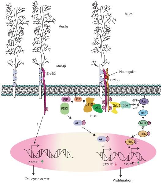
Mucins are high-molecular-weight glycoproteins and are implicated in diverse biological functions. MUC4, a member of transmembrane mucin family, is expressed in airway epithelial cells and body fluids like saliva, tear film, ear fluid, and breast milk. In addition to its normal expression, an aberrant expression of MUC4 has been reported in a variety of carcinomas. Among various potential domains of MUC4, epidermal growth factor (EGF) -like domains are hypothesized to interact with and activate the ErbB2 receptors, suggesting an intramembrane-growth factor function for MUC4. The heavily glycosylated tandem repeat domain provides the structural rigidity to the extended extracellular region. MUC4, by virtue of its extended structure, serves as a barrier for some cell-cell and cell-extracellular matrix interactions and as a potential reservoir for certain growth factors. An intricate relationship between MUC4 and growth factor signaling is also reflected in the transcriptional regulation of MUC4. The MUC4 promoter has binding sites for different transcription factors, which are responsible for the regulation of its expression in different tissues. The interferon-gamma, retinoic acid, and transforming growth factor-beta signaling pathways regulate MUC4 expression in a partially interdependent manner. Taken together, all of these features of MUC4 strongly support its role as a potential candidate for diagnostic and therapeutic applications in cancer and other diseases.


Figures

Figure 1
Figure 1
MUC4 structure. A) MUC4 gene is encoded by 26 exons (E1–E26). E1 exon codes for amino-terminal of the protein. E2 is the largest. E2 is also polymorphic and codes for the central domain. Exons E3–E26 code for the carboxyl-terminal of the MUC4 protein, which includes various domains present in MUC4-like NIDO, AMOP, vWD, transmembrane region, and the cytoplasmic tail. B) MUC4 protein is divided in three regions: the N- terminal (NT), the C region (central domain), and the CT region. The C-terminal region codes for 12 domains (CT1–CT12). Different domains present in MUC4 protein are the central large tandem repeat domain, NIDO, AMOP, vWD, and 3 carboxyl-terminal located EGF domains. The MUC4 protein is hypothesized to be cleaved at GDPH, proteolytic site, generating two subunits: MUC4α and MUC4β. MUC4α is a mucin-like subunit that is heavily glycosylated, and MUC4β is a growth factor like subunit due to presence of EGF-like domains. MUC4 is anchored to the cell surface by the transmembrane region. MUC4 has a short cytoplasmic tail of 22 amino acids.
Figure 2
Figure 2
Evolution of the MUC4 mucin. A) Alignment of amino acid sequences of various protein domains of MUC4 from human, dog, mouse, rat, and chicken. The domains were defined by a simple modular architecture research tool (SMART) domain search and a Prosite search. The alignment was performed by using the ClustalW program of the European Bioinformatic Institute (EBI). “*” Indicates residues are identical in all sequences in the alignment. “:” Indicates conserved substitutions (within the amino acid subgroups). “.” Indicates semiconserved substitutions. According to the standard coloring scheme of EBI, red indicates small and hydrophobic residues. Blue and magenta indicate acidic and basic residues, respectively. Green represents amino acids containing hydroxyl and amine groups, while gray represents other residues. B) Phylogenetic tree of MUC4 orthologues from different species. Evolutionary relatedness of human and dog sequences and rat and mouse sequences was observed by sequence-based alignment. The phylogenetic tree was generated by the EBI ClustalW program by using complete protein sequences of different MUC4 orthologues. The branch lengths indicate the amount of evolutionary change. C) Evolution scheme of different domains of the MUC4 mucin. Each rectangle in the tandem repeat region represents a 16 aa motif that is repeated 146–500 times. The tandem repeat region probably evolved from duplications of Ser-Thr rich regions in ancestral protein A. The NIDO and EGF-like domains evolved from ancestral protein B, which is a common progenitor to the nidogen protein. Similarly, AMOP and vWD domains evolved from ancestral protein C, a common precursor to Susd2 protein. Domain structures of MUC4, nidogen, and Susd2 were analyzed using SMART. Nidogen_G2 = a β-barrel domain of nidogen; EGF_3 = EGF-like domain; THYROG = throglobulin type-1 domain; LDLRB = low-density lipoprotein-receptor class B; SMB_2 = somatomedin B domain; SUSHI = Sushi/SCP/SCR domain.
Figure 2
Figure 2
Evolution of the MUC4 mucin. A) Alignment of amino acid sequences of various protein domains of MUC4 from human, dog, mouse, rat, and chicken. The domains were defined by a simple modular architecture research tool (SMART) domain search and a Prosite search. The alignment was performed by using the ClustalW program of the European Bioinformatic Institute (EBI). “*” Indicates residues are identical in all sequences in the alignment. “:” Indicates conserved substitutions (within the amino acid subgroups). “.” Indicates semiconserved substitutions. According to the standard coloring scheme of EBI, red indicates small and hydrophobic residues. Blue and magenta indicate acidic and basic residues, respectively. Green represents amino acids containing hydroxyl and amine groups, while gray represents other residues. B) Phylogenetic tree of MUC4 orthologues from different species. Evolutionary relatedness of human and dog sequences and rat and mouse sequences was observed by sequence-based alignment. The phylogenetic tree was generated by the EBI ClustalW program by using complete protein sequences of different MUC4 orthologues. The branch lengths indicate the amount of evolutionary change. C) Evolution scheme of different domains of the MUC4 mucin. Each rectangle in the tandem repeat region represents a 16 aa motif that is repeated 146–500 times. The tandem repeat region probably evolved from duplications of Ser-Thr rich regions in ancestral protein A. The NIDO and EGF-like domains evolved from ancestral protein B, which is a common progenitor to the nidogen protein. Similarly, AMOP and vWD domains evolved from ancestral protein C, a common precursor to Susd2 protein. Domain structures of MUC4, nidogen, and Susd2 were analyzed using SMART. Nidogen_G2 = a β-barrel domain of nidogen; EGF_3 = EGF-like domain; THYROG = throglobulin type-1 domain; LDLRB = low-density lipoprotein-receptor class B; SMB_2 = somatomedin B domain; SUSHI = Sushi/SCP/SCR domain.
Figure 3
Figure 3
rMuc4 and ErbB signaling. The interaction of rMuc4 with ErbB2 induces its limited phosphorylation and an up-regulation of p27Kip1, leading to cell cycle arrest. In the presence of neuregulin, rMuc4 is engaged in a quad-complex with ErbB2, ErbB3, and neuregulin and potentiate phosphorylation of ErbB2 leading to an Akt-mediated survival (via down-regulation of p27Kip1) and an ERK-mediated proliferative response. MEK = MAP-ERK kinase; PDK = phosphoinositide-dependent kinase; PIP = phosphatidylinositol phosphate.
Figure 4
Figure 4
Regulation of MUC4 expression. A) Schematic of MUC4 promoter depicting the presence of various transcription factor binding sites AP-1, growth response element (GRE), hepatocyte nuclear factor 1α (HNF1α), STAT, retinoic acid receptor (RAR), SMAD, nuclear factor-κB (κB), retinoid receptor (RXR), and Sp1. The expression of MUC4 can be driven by an active proximal and a distal promoter region. The proximal promoter does not contain a TATA box, while it is present in the distal promoter region. B) Expression of MUC4 is transcriptionally regulated by a variety of growth factors, cytokines, and other components (IFNγ, RA, TGFβ, interleukins, and bile acids). IFNγ-induced expression of MUC4 is mediated by a novel mechanism that involves an up-regulation of STAT1. Retinoic acid-induced expression of MUC4 is mediated by TGFβ2. Induction of MUC4 expression by TGFβ occurs via both SMAD-dependent and -independent pathways. TGFβ-stimulation activates SMAD2/3, which in complex with SMAD4 gets translocated to the nucleus and facilitates the transcription of MUC4. In addition, TGFβ can drive MUC4 expression via MAPK, PI3K, and PKA pathways. Interleukins are known to regulate MUC4 expression via the STAT6 pathway in airway epithelial cells. Bile acids up-regulate MUC4 expression in esophageal cancer cells by involving the HNF1α transcription factor. RBP = retinoic acid binding protein; TCDC = taurochenodeoxycholate; TDC = taurodeoxycholate.
Figure 5
Figure 5
Multiple roles of MUC4 mucin in cancer development. MUC4 is engaged in complex formation with ErbB2 and/or ErbB3 (in presence of neuregulin) and initiates and/or potentiates downstream signaling and facilitates cell proliferation and cell survival. The large sized extracellular domain of MUC4 disrupts cell-cell and cell-extracellular matrix interactions via steric hindrance. MUC4 also increases cell motility via yet unknown mechanisms. The sialyl epitopes present on the heavily glycosylated tandem repeat domain of MUC4 may facilitate trans-interactions by binding to selectins or yet uncharacterized ligands on endothelial cells. The presence of MUC4 on the surface of the tumor cells can mask the surface epitopes to the cytotoxic immune cells such as cytotoxic-T lymphocytes or NK cells and, hence, escape from immune response.

References

    1. Hollingsworth MA, Swanson BJ. Mucins in cancer: protection and control of the cell surface. Nat. Rev. Cancer. 2004;4:45–60. - PubMed
    1. Moniaux N, Escande F, Porchet N, Aubert JP, Batra SK. Structural organization and classification of the human mucin genes. Front. Biosci. 2001;6:D1192–1206. - PubMed
    1. Satoh S, Hinoda Y, Hayashi T, Burdick MD, Imai K, Hollingsworth MA. Enhancement of metastatic properties of pancreatic cancer cells by MUC1 gene encoding an anti-adhesion molecule. Int. J. Cancer. 2000;88:507–518. - PubMed
    1. Wesseling J, van der Valk SW, Vos HL, Sonnenberg A, Hilkens J. Episialin (MUC1) overexpression inhibits integrin-mediated cell adhesion to extracellular matrix components. J. Cell Biol. 1995;129:255–265. - PMC - PubMed
    1. Strous GJ, Dekker J. Mucin-type glycoproteins. Crit. Rev. Biochem. Mol. Biol. 1992;27:57–92. - PubMed

Publication types