Comparative Study
. 2008 Mar;102(1):160-70.
doi: 10.1093/toxsci/kfm283. Epub 2007 Nov 17.

The aryl hydrocarbon receptor affects distinct tissue compartments during ontogeny of the immune system

Affiliations
Comparative Study

The aryl hydrocarbon receptor affects distinct tissue compartments during ontogeny of the immune system

Jason P Hogaboam et al. Toxicol Sci. 2008 Mar.

Abstract

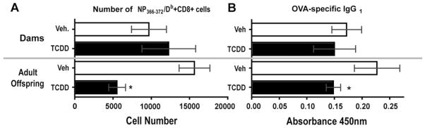
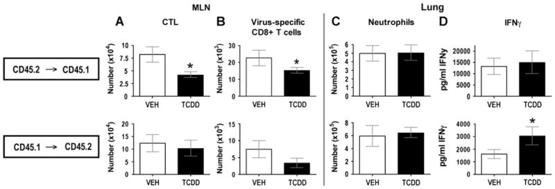
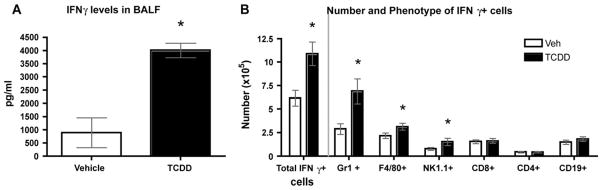
There is growing evidence that prenatal and early postnatal environmental factors influence the development and programming of the immune system, causing long-lasting negative health consequences. The aryl hydrocarbon receptor (AhR) is an important modulator of the development and function of the immune system; however, the mechanism is poorly understood. Exposure to the AhR agonist 2,3,7,8-tetrachlorodibenzo-p-dioxin throughout gestation and during lactation yields adult offspring with persistent defects in their immune response to influenza virus. These functional alterations include suppressed lymphocyte responses and increased inflammation in the infected lung despite normal cellularity and anatomical development of lymphoid organs. The studies presented here were conducted to determine the critical period during immune ontogeny that is particularly sensitive to inappropriate AhR activation. We also investigated the contribution of AhR-mediated events within and extrinsic to hematopoietic cells. Our findings show that AhR activation alters different elements of the immune system at different times during development by affecting different tissue targets. In particular, diminished T-cell responses arise due to deregulated events within bone marrow-derived cells. In contrast, increased interferon gamma levels in the infected lung result from AhR-regulated events extrinsic to bone marrow-derived cells, and require AhR agonist exposure during early gestation. The persistence of AhR activation induced immune modulation was also compared, revealing that AhR activation causes long-lasting functional alterations in the developing immune system, whereas the impact on the mature immune system is transient.


Figures

FIG. 1
FIG. 1
AhR activation during the ontogeny of the immune system causes long-lasting functional deregulation. Responses to influenza virus and OVA were compared in vehicle- and TCDD-treated dams and their developmentally exposed adult offspring. Antigen was administered to dams 8 weeks after their last dose of TCDD (i.e., when their offspring had reached maturity). Treated dams were of the same age, and data from TCDD-exposed offspring are compared with age-matched vehicle-exposed offspring. (A) Graphs depict the average number of virus-specific CD8+ cells from vehicle- and TCDD-treated dams and their adult offspring 9 days after infection (i.n.) with 120 HAU influenza A virus (×31). MLN cells were stained with fluorescently labeled MHC class I molecules complexed with NP366–372 and antibodies against CD8 and analyzed by flow cytometry (B) The average level of OVA-specific IgG1 in the plasma was determined in a separate group of treated dams and their offspring, which had been sensitized and challenged with OVA as described in the “Materials and Methods.” OVA-specific IgG1 was measured by isotype-specific ELISA. Absorbance (optical density) readings represent those from a 1:2.6 × 10⁶ dilution of plasma, which was within the linear range of the dilution series. Findings were similar when plasma OVA-specific IgE levels were examined (data not shown). For these studies, there were three to four dams or six to eight pups per treatment group. Error bars indicate SEM. An * indicates a significant difference from vehicle-exposed controls (p ≤ 0.05).
FIG. 2
FIG. 2
Dosing strategy of timed pregnant mice for developmental exposure to TCDD. Impregnated mice were treated with vehicle or 1 μg/kg TCDD on gd 0, gd 7, and gd 14 and/or 2 days after parturition (filled diamond). A subset of these doses was administered to separate groups of dams, indicated by the blocks underneath the dosing strategy. Mice that received only gestational exposure to TCDD were cross-fostered to untreated dams 2 days after birth. Offspring that received only lactational exposure were born to untreated dams and cross-fostered to treated dams that had received TCDD on gd 0, gd 7, gd 14 and 2 days after parturition. This cross-fostering insured that treated dams were exposed to the same amount of TCDD. Critical events during ontogeny of the vertebrate immune system are represented above the dosing scheme.
FIG. 3
FIG. 3
Developmental exposure to TCDD throughout all of gestation and then via lactation results in impaired immune function. Developmentally exposed adult mice (6–8 weeks of age) were treated as described in Figure 2, and the following abbreviations are used: exposure during all of gestation and via lactation (G + L), gestation only (G only), lactation only (L only), or late gestation and lactation (late G + L). Data from TCDD-exposed offspring are compared with age-matched vehicle-exposed offspring that were infected at the same time. BAL cells and BAL fluid were collected 7 days after infection with influenza virus, whereas MLN cells were collected on day 9. All graphs depict the average fold change relative to infected adult offspring of control dams (TCDD/Veh), in which a value of 1 means that there was no difference between the groups. (A) The number of NP366–372/Db+CD8+ cells was determined by flow cytometry. (B) The average number of neutrophils in the lung was determined by differential cell counting. (C) The average amount of IFN-γ in lung lavage fluid was determined by ELISA. There were five to eight female offspring per treatment group at each point in time, and each infected mouse was from a different exposed dam. Error bars indicate SEM. An * indicates a significant difference from infected offspring of vehicle-exposed dams (p ≤ 0.05).
FIG. 4
FIG. 4
Generation of bone marrow chimeras is not affected by maternal TCDD exposure. (A) Experimental design. CD45.2 → CD45.1 chimeras were generated by transferring bone marrow cells (BMC) from age-matched female C57Bl/6 CD45.2 mice that were developmentally exposed to TCDD or vehicle (as described in Fig. 2, G + L) into lethally irradiated (but otherwise untreated) adult B6.CD45.1 congenic mice. CD45.1 → CD45.2 chimeras were created by transferring BMC from untreated B6.CD45.1 congenic female mice into lethally irradiated, age-matched adult C57Bl/6 CD45.2 mice that had been developmentally exposed to TCDD or vehicle (as described in Fig. 2, G + L). (B) Representative histograms show that 6–8 weeks after reconstitution, bone marrow cells in the recipient mice are derived from the donor. The number beside each histogram shows the average percentage of cells in bone marrow from each treatment group (± SEM). There were no significant differences in chimerization among the groups.
FIG. 5
FIG. 5
AhR-mediated events within hematopoietic cells underlie suppressed response of virus-specific CD8+ T cells, but not enhanced innate responses to infection. Reciprocal chimeras, generated as described in Figure 4, were challenged with influenza virus 6–8 weeks later. (A, B) MLN cells were obtained 9 days after x31 infection. Graphs depict the average number of (A) CTLe (CD44hiCD62LloCD8+ cells) and (B) NP366–372/Db+CD8+ cells in each group of chimeras. (C, D) Airway cells and BAL fluid were collected on day 7 following infection with x31, and graphs show (C) the average number of neutrophils and (D) level of IFN-γ in BAL fluid. Fluorescently labeled antibodies against CD45.1 and CD45.2 were used to validate chimerization, and to distinguish chimeric cells from residual endogenous cells (see Fig. 4B). Results are representative of at least two separate experiments. In each experiment, there were five to six female mice in each treatment group at each point in time. Error bars indicate SEM. An * indicates a significant difference from the vehicle-treatment group (p ≤ 0.05).
FIG. 6
FIG. 6
Excess IFN-γ in lungs of infected mice that were developmentally exposed to TCDD comes primarily from phagocytic cells. Lung-derived immune cells and lung lavage fluid from vehicle and TCDD developmentally exposed offspring (8 weeks of age; G + L, Fig. 2) were collected 7 days after infection with influenza virus (x31). (A) Average level of IFN-γ in the BAL fluid, as measured by ELISA. (B) Bar graphs depict the number and phenotype of IFN-γ+ cells in the lung. Lung-derived leukocytes were restimulated in vitro, stained with fluorescently labeled antibodies and analyzed by flow cytometry as described in the “Materials and Methods.” There were 7–10 female mice in each treatment group, and each infected mouse was from a different exposed dam. Error bars indicate SEM. An * indicates a significant difference from infected offspring of vehicle-exposed dams (p ≤ 0.05). Data are representative of two separate experiments.

References

    1. Allan L, Mann K, Matulka R, Ryu H, Schlezinger J, Sherr D. Bone marrow stromal-B cell interactions in polycyclic aromatic hydrocarbon-induced pro/pre-B cell apoptosis. Toxicol Sci. 2003;76:357–365. - PubMed
    1. Andersson P, Ridderstad A, McGuire J, Pettersson S, Poellinger L, Hanberg A. A constitutively active aryl hydrocarbon receptor causes loss of peritoneal B1 cells. Biochem Biophys Res Commun. 2003;302:236–241. - PubMed
    1. Birnbaum L. Distribution and excretion of 2,3,7,8-tetrachlorodibenzo-p-dioxin in congenic strains of mice which differ at the Ah locus. Drug Metab Dispos. 1986;14:34–40. - PubMed
    1. Camacho I, Nagarkatti M, Nagarkatti P. Evidence for induction of apoptosis in T cells from murine fetal thymus following perinatal exposure to 2,3,7,8-tetrachlorodibenzo-p-dioxin. Toxicol Sci. 2004;78:96–106. - PubMed
    1. Camacho I, Singh N, Hegde V, Nagarkatti M, Nagarkatti P. Treatment of mice with 2,3,7,8-tetrachlorodibenzo-p-dioxin leads to aryl hydrocarbon receptor-dependent nuclear translocation of NF-kappa B and expression of fas ligand in thymic stromal cells and consequent T cell apoptosis. J Immunol. 2005;175:90–103. - PubMed

Publication types

MeSH terms

Substances