Comparative Study
. 2008 Feb;76(2):542-50.
doi: 10.1128/IAI.00952-07. Epub 2007 Nov 19.

Genomic comparison of virulent Rickettsia rickettsii Sheila Smith and avirulent Rickettsia rickettsii Iowa

Affiliations
Comparative Study

Genomic comparison of virulent Rickettsia rickettsii Sheila Smith and avirulent Rickettsia rickettsii Iowa

Damon W Ellison et al. Infect Immun. 2008 Feb.

Abstract

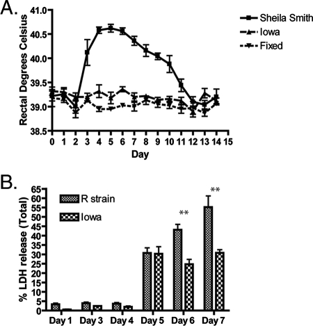
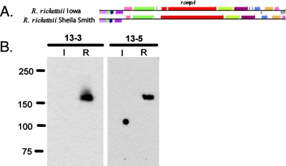
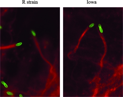
Rickettsia rickettsii is an obligate intracellular pathogen that is the causative agent of Rocky Mountain spotted fever. To identify genes involved in the virulence of R. rickettsii, the genome of an avirulent strain, R. rickettsii Iowa, was sequenced and compared to the genome of the virulent strain R. rickettsii Sheila Smith. R. rickettsii Iowa is avirulent in a guinea pig model of infection and displays altered plaque morphology with decreased lysis of infected host cells. Comparison of the two genomes revealed that R. rickettsii Iowa and R. rickettsii Sheila Smith share a high degree of sequence identity. A whole-genome alignment comparing R. rickettsii Iowa to R. rickettsii Sheila Smith revealed a total of 143 deletions for the two strains. A subsequent single-nucleotide polymorphism (SNP) analysis comparing Iowa to Sheila Smith revealed 492 SNPs for the two genomes. One of the deletions in R. rickettsii Iowa truncates rompA, encoding a major surface antigen (rickettsial outer membrane protein A [rOmpA]) and member of the autotransporter family, 660 bp from the start of translation. Immunoblotting and immunofluorescence confirmed the absence of rOmpA from R. rickettsii Iowa. In addition, R. rickettsii Iowa is defective in the processing of rOmpB, an autotransporter and also a major surface antigen of spotted fever group rickettsiae. Disruption of rompA and the defect in rOmpB processing are most likely factors that contribute to the avirulence of R. rickettsii Iowa. Genomic differences between the two strains do not significantly alter gene expression as analysis of microarrays revealed only four differences in gene expression between R. rickettsii Iowa and R. rickettsii strain R. Although R. rickettsii Iowa does not cause apparent disease, infection of guinea pigs with this strain confers protection against subsequent challenge with the virulent strain R. rickettsii Sheila Smith.


Figures

FIG. 1.
FIG. 1.
R. rickettsii Iowa virulence and the ability of R. rickettsii Iowa to lyse Vero cells are attenuated. (A) Female Hartley guinea pigs were infected with 1,000 PFU of R. rickettsii Sheila Smith or R. rickettsii Iowa or inoculated with paraformaldehyde-fixed rickettsiae (Fixed), and their temperatures were monitored for 14 days postinfection. (B) Monolayers of Vero cells grown in 96-well plates were infected with either R. rickettsii strain R or R. rickettsii Iowa, and the percentages of LDH released into the media were determined on days 1, 3, 4, 5, 6, and 7 postinfection. Statistically significant differences (P < 0.001) are indicated above the bars (two asterisks) and were determined by performing a two-way analysis of variance for the relative fluorescence units using GraphPad Prism software (n = 5).
FIG. 2.
FIG. 2.
Phylogram showing that R. rickettsii Sheila Smith and R. rickettsii Iowa are closely related. The following concatenated sequences of Rickettsia species were used for the analysis: glt, gyrB, ompB, recA, and sca4. R. bellii was used as an outgroup. The tree was constructed with Clustal V and the neighbor-joining method with 10,000 bootstrap replicates. A number at a node is the percentage of bootstrap replicates that supported the branching pattern to the right. The scale bar for the branch lengths indicates the number of substitutions per site.
FIG. 3.
FIG. 3.
Circular diagram of the R. rickettsii Iowa genome. The outer circle is an alignment of each ORF identified in R. rickettsii Iowa compared to ORFs in R. rickettsii Sheila Smith. ORFs that share an E value cutoff of ≥1e10−10 are gray, while ORFs that lack R. rickettsii Iowa homology are black. The second and third circles are the predicted ORFs in R. rickettsii Iowa and the strand of DNA that encodes them (orange, positive strand; blue, negative strand). The fourth circle shows the locations of RNA-encoding regions; tRNAs are green, and rRNAs are red. The fifth circle shows all of the ORFs color coded by functional category, as indicated at the bottom. The sixth circle shows GC skew, and the seventh circle shows the G+C content with a sliding 20-kb window.
FIG. 4.
FIG. 4.
Evidence of genomic reduction in R. rickettsii Sheila Smith: graphic representation of the genomic region deleted from R. rickettsii Sheila Smith. The region deleted from R. rickettsii Sheila Smith is indicated by black arrows. CoA, coenzyme A.
FIG. 5.
FIG. 5.
rOmpA is disrupted in R. rickettsii Iowa. (A) Graphic representation of the disrupted rompA gene in R. rickettsii Iowa. (B) Equal amounts of R. rickettsii Iowa (lanes I) and R. rickettsii strain R (lanes R) were loaded and run on a 10% sodium dodecyl sulfate-polyacrylamide gel electrophoresis gel and transferred to a nitrocellulose membrane. rOmpA was detected using monoclonal antibodies 13-3 and 13-5 (2).
FIG. 6.
FIG. 6.
R. rickettsii Iowa shows normal actin tail formation. Monolayers of Vero cells were infected with either R. rickettsii Iowa or R. rickettsii strain R and allowed to grow for 1 day. R. rickettsii was detected using monoclonal antibody 13-2 (1/100), while F-actin was labeled with rhodamine phalloidin (10 U/ml).
FIG. 7.
FIG. 7.
SNPs present in R. rickettsii Iowa and R. rickettsii Sheila Smith. The numbers of SNPs found in specific ORFs are indicated on the x axis. The total numbers of SNPs/ORF are indicated on the y axis.

References

    1. Anacker, R. L., R. H. List, R. E. Mann, and D. L. Wiedbrauk. 1986. Antigenic heterogeneity in high- and low-virulence strains of Rickettsia rickettsii revealed by monoclonal antibodies. Infect. Immun. 51653-660. - PMC - PubMed
    1. Anacker, R. L., R. E. Mann, and C. Gonzales. 1987. Reactivity of monoclonal antibodies to Rickettsia rickettsii with spotted fever and typhus group rickettsiae. J. Clin. Microbiol. 25167-171. - PMC - PubMed
    1. Anacker, R. L., T. F. McCaul, W. Burgdorfer, and R. K. Gerloff. 1980. Properties of selected rickettsiae of the spotted fever group. Infect. Immun. 27468-474. - PMC - PubMed
    1. Anacker, R. L., R. N. Philip, J. C. Williams, R. H. List, and R. E. Mann. 1984. Biochemical and immunochemical analysis of Rickettsia rickettsii strains of various degrees of virulence. Infect. Immun. 44559-564. - PMC - PubMed
    1. Andersson, S. G., and C. G. Kurland. 1998. Reductive evolution of resident genomes. Trends Microbiol. 6263-268. - PubMed

Publication types

MeSH terms

Associated data