. 2008 Feb;76(2):515-22.
doi: 10.1128/IAI.01064-07. Epub 2007 Nov 19.

Caspase-1 contributes to Chlamydia trachomatis-induced upper urogenital tract inflammatory pathologies without affecting the course of infection

Affiliations

Caspase-1 contributes to Chlamydia trachomatis-induced upper urogenital tract inflammatory pathologies without affecting the course of infection

Wen Cheng et al. Infect Immun. 2008 Feb.

Abstract

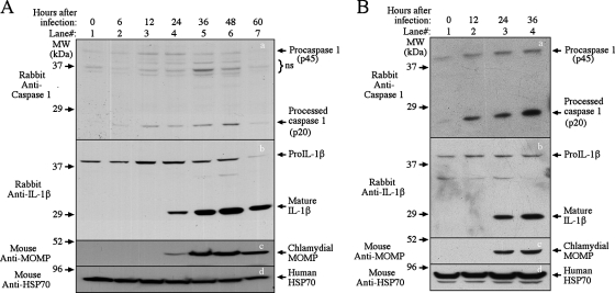
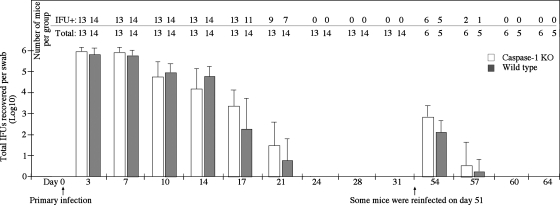
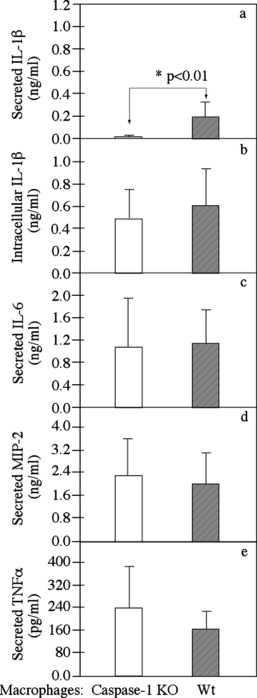
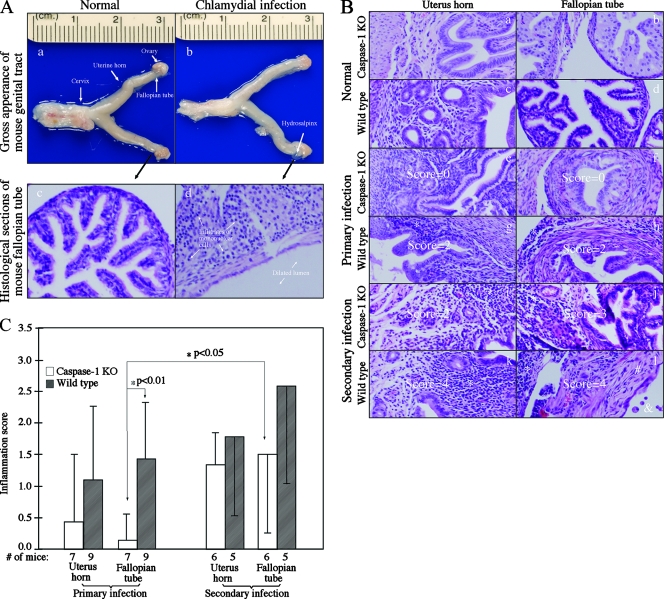
Chlamydia trachomatis infection induces inflammatory pathologies in the upper genital tract, potentially leading to ectopic pregnancy and infertility in the affected women. Caspase-1 is required for processing and release of the inflammatory cytokines interleukin-1beta (IL-1beta), IL-18, and possibly IL-33. In the present study, we evaluated the role of caspase-1 in chlamydial infection and pathogenesis. Although chlamydial infection induced caspase-1 activation and processing of IL-1beta, mice competent and mice deficient in caspase-1 experienced similar courses of chlamydial infection in their urogenital tracts, suggesting that Chlamydia-activated caspase-1 did not play a significant role in resolution of chlamydial infection. However, when genital tract tissue pathologies were examined, the caspase-1-deficient mice displayed much reduced inflammatory damage. The reduction in inflammation was most obvious in the fallopian tube tissue. These observations demonstrated that although caspase-1 is not required for controlling chlamydial infection, caspase-1-mediated responses can exacerbate the Chlamydia-induced inflammatory pathologies in the upper genital tract, suggesting that the host caspase-1 may be targeted for selectively attenuating chlamydial pathogenicity without affecting the host defense against chlamydial infection.


Figures

FIG. 1.
FIG. 1.
Activation of caspase-1 by chlamydial infection. HeLa cells infected with C. trachomatis (A) or C. muridarum (MoPn) (B) were harvested at various time points after infection as indicated at the top in order to monitor caspase-1 (panels a) and IL-1β (panels b) processing in a Western blot. The anti-caspase-1 and IL-1β antibodies indicated on the left detected both the pro and mature forms of caspase-1 and IL-1β, as indicated on the right. A mouse anti-chlamydial major outer membrane protein (MOMP) monoclonal antibody was used to monitor chlamydial infection (panels c), and an anti-mammalian heat shock protein 70 (HSP70) antibody was used to monitor total protein loading (panels d). The sample harvested at zero time was normal HeLa cells not infected with chlamydiae (lane 1). Note that both caspase-1 processing and IL-1β processing were induced by chlamydial infection. MW, molecular mass; ns, not significant.
FIG. 2.
FIG. 2.
Effect of caspase-1 deficiency on live chlamydia shedding following chlamydial infection. Mice deficient (open bars) or competent (filled bars) for caspase-1 were infected intravaginally with C. muridarum, and vaginal swabs were taken during the course of infection as indicated on the x axis to determine the number of live organisms (expressed in IFUs) shed from the urogenital tract. The number of IFU obtained from each swab was converted into a log10 value, and the log10 IFU values were used to calculate the mean and standard deviation for each mouse group at each time point, as shown on the y axis. At the start of the experiment the caspase-1 KO group contained 13 mice, while the wild-type (wt) group contained 14 mice. The number of mice with detectable IFUs at each time point is indicated above the horizontal line at the top. On day 51 after the primary infection, six and five mice in the caspase-1 KO and wild-type groups, respectively, were reinfected with C. muridarum. All mice were sacrificed on day 80 after the primary infection. The log10 IFU values for the KO and wild-type groups at each time point were analyzed using a two-tailed Student t test, and no statistically significant differences were found. Note that the course of infection was dramatically shortened following the secondary infection.
FIG. 3.
FIG. 3.
Effect of caspase-1 deficiency on cytokine production and secretion by mouse Mφs after Chlamydia infection. Peritoneal Mφs harvested from either caspase-1 KO (open bars) or wild-type (filled bars) mice were infected with chlamydiae for 24 h in 48-well plates. The intracellular IL-1β (b), secreted IL-1β (a), IL-6 (c), macrophage inflammatory protein 2 (MIP-2) (mouse IL-8 homolog) (d), and tumor necrosis factor alpha (TNFα) (panel e) levels were measured using a commercially available ELISA kit. The data were obtained using seven to nine mice in each group and were expressed in ng or pg per ml of culture supernatant; the bars indicate means, and the error bars indicate standard deviations. The asterisk indicates a statistically significant difference (P < 0.01) in the level of secreted IL-1β between the caspase-1 KO and wild-type Mφs (both infected with chlamydiae).
FIG. 4.
FIG. 4.
Effect of caspase-1 deficiency on the development of inflammatory pathologies in the mouse urogenital tract following chlamydial infection. (A) When urogenital tract tissues from wild-type NOD mice with (panels b and d) or without (panels a and c) a chlamydial infection were examined at the level of gross appearance (panels a and b) and with a microscope (panels c and d), obvious inflammatory pathologies were noted in the samples from the infected mouse but not in the samples from the normal mouse, and these pathologies included hydrosalpinx formation (panel b, white arrow), extensive infiltration of mononuclear cells (panel d, arrows), and fallopian tube luminal dilation (panel d, arrows). Different types of tissues in a NOD mouse urogenital tract with a normal gross appearance are indicated in panel a (arrows). (B) Mouse genital tract tissues were sectioned and analyzed using a microscope after H&E staining. A representative image of a histological section from either the uterine horn (panels a, c, e, g, I, and k) or fallopian tube (panels b, d, f, h, j, and l) tissues is shown for each group of mice with or without a primary or secondary infection. The asterisks indicate inflammatory cell infiltration, the number sign indicates fibrosis, and the ampersand indicates luminal dilation. Scores based on the severity of inflammatory infiltration are indicated in some of the images. Note that chlamydial infection induced more severe inflammatory changes in wild-type mice. (C) Inflammation scores assigned to individual mice were used to calculate the means (bars) and standard errors (error bars) for different groups. The various tissue and mouse groups are indicated on the x axis. The open bars indicate tissue samples from caspase-1 KO mice, while the filled bars indicate tissue samples from wild-type mice. An analysis of variance test was used to analyze differences among different groups, and a two-tailed Student t test was used to analyze differences between the caspase-1 KO and wild-type groups. There was a highly significant difference in inflammation scores (P < 0.01, Student t test) between the caspase-1 KO and wild-type fallopian tissues, and there was a significant difference in the caspase-1 KO fallopian tube tissue inflammation (P < 0.05, Student t test) between the primary and secondary infections.

References

    1. Barteneva, N., I. Theodor, E. M. Peterson, and L. M. de la Maza. 1996. Role of neutrophils in controlling early stages of a Chlamydia trachomatis infection. Infect. Immun. 644830-4833. - PMC - PubMed
    1. Bauwens, J. E., H. Orlander, M. P. Gomez, M. Lampe, S. Morse, W. E. Stamm, R. Cone, R. Ashley, P. Swenson, and K. K. Holmes. 2002. Epidemic Lymphogranuloma venereum during epidemics of crack cocaine use and HIV infection in the Bahamas. Sex. Transm. Dis. 29253-259. - PubMed
    1. Bilenki, L., S. Wang, J. Yang, Y. Fan, A. G. Joyee, and X. Yang. 2005. NK T cell activation promotes Chlamydia trachomatis infection in vivo. J. Immunol. 1753197-3206. - PubMed
    1. Boraschi, D., and A. Tagliabue. 2006. The interleukin-1 receptor family. Vitam. Horm. 74229-254. - PubMed
    1. Carriere, V., L. Roussel, N. Ortega, D. A. Lacorre, L. Americh, L. Aguilar, G. Bouche, and J. P. Girard. 2007. IL-33, the IL-1-like cytokine ligand for ST2 receptor, is a chromatin-associated nuclear factor in vivo. Proc. Natl. Acad. Sci. USA 104282-287. - PMC - PubMed

Publication types