. 2008 Feb;28(3):1182-94.
doi: 10.1128/MCB.01500-07. Epub 2007 Nov 19.

SLIP1, a factor required for activation of histone mRNA translation by the stem-loop binding protein

Affiliations

SLIP1, a factor required for activation of histone mRNA translation by the stem-loop binding protein

Nihal G Cakmakci et al. Mol Cell Biol. 2008 Feb.

Abstract

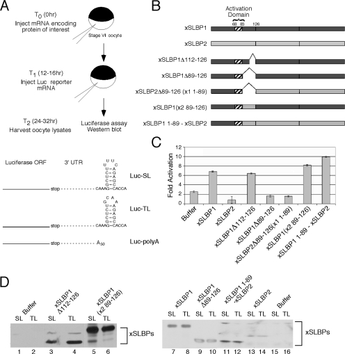
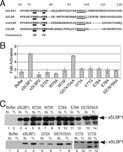
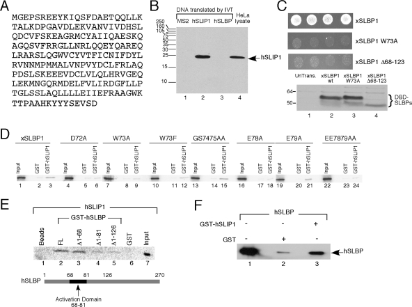
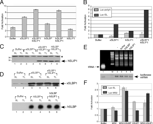
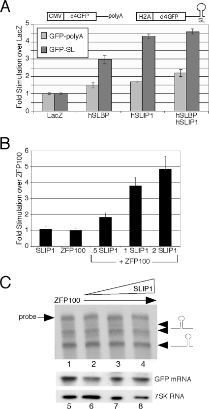
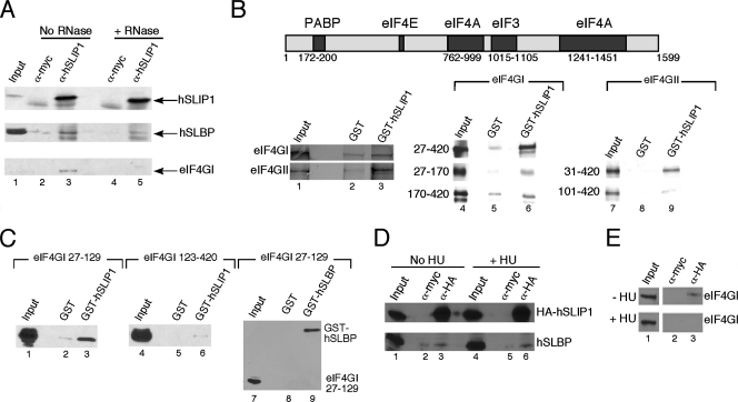
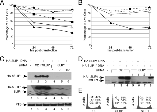
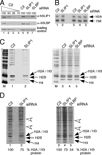
Replication-dependent histone mRNAs are the only eukaryotic cellular mRNAs that are not polyadenylated, ending instead in a conserved stem-loop. The 3' end of histone mRNA is required for histone mRNA translation, as is the stem-loop binding protein (SLBP), which binds the 3' end of histone mRNA. We have identified five conserved residues in a 15-amino-acid region in the amino-terminal portion of SLBP, each of which is required for translation. Using a yeast two-hybrid screen, we identified a novel protein, SLBP-interacting protein 1 (SLIP1), that specifically interacts with this region. Mutations in any of the residues required for translation reduces SLIP1 binding to SLBP. The expression of SLIP1 in Xenopus oocytes together with human SLBP stimulates translation of a reporter mRNA ending in the stem-loop but not a reporter with a poly(A) tail. The expression of SLIP1 in HeLa cells also stimulates the expression of a green fluorescent protein reporter mRNA ending in a stem-loop. RNA interference-mediated downregulation of endogenous SLIP1 reduces the rate of translation of endogenous histone mRNA and also reduces cell viability. SLIP1 may function by bridging the 3' end of the histone mRNA with the 5' end of the mRNA, similar to the mechanism of translation of polyadenylated mRNAs.


Figures

FIG. 1.
FIG. 1.
Analysis of activity of deletion mutants of xSLBP1 in Xenopus oocytes. (A) Schematic of Xenopus oocyte system used as an in vivo translation assay (24). The structures of the luciferase reporters Luc-SL, Luc-TL, and Luc-poly(A) are shown. Abbreviations: ORF, open reading frame; UTR, untranslated region. (B) The structure of the mutant proteins. The central domain of the xSLBPs contains the RBD, beginning with amino acid 126, and the translation activation domain of xSLBP1 is indicated (amino acids 68 to 83). (C) Synthetic mRNAs encoding each of these proteins were injected into Xenopus oocytes, and 16 h later the Luc-SL or Luc-TL mRNAs were injected. Sixteen hours later, the oocytes were harvested and luciferase activity was measured. Results are normalized to the Luc-TL activity. (D) The levels of the expressed proteins in the experiment shown in panel C were analyzed by Western blotting using anti-xSLBP1 antibody (which cross-reacts weakly with xSLBP2 (lanes 13 and 14) to demonstrate the expression of the mutant proteins.
FIG. 2.
FIG. 2.
Identification of amino acids in the translation activation region of SLBP required for activity. (A) The sequences of xSLBP1, hSLBP, sea urchin SLBP (suSLBP), and Ciona SLBP (cSLBP) are compared in the region just before the RBD. The numbering is for xSLBP1. The thickly underlined region(s) within 72 to 79 is the consensus core translation activation region, and the thinly underlined region from 95 to 99 is the cyclin binding site required for SLBP degradation (38). (B) Wild-type xSLBP1, xSLBP2, or the indicated mutants of xSLBP1 were expressed in Xenopus oocytes by injection of synthetic capped mRNA as described in Fig. 1A. Results are expressed relative to the expression of the Luc-TL mRNA, which was set at 1. Oocytes injected with buffer served as a control. They have activity higher than 1 because of the endogenous xSLBP1 in the oocyte. For each point, 12 oocytes were pooled. The results are an average of three independent experiments using oocytes from different frogs. (C) Protein from the equivalent of 0.5 oocyte that had been injected with either Luc-SL or Luc-TL reporter in Fig. 2B was resolved on 12% SDS-PAGE. The amounts of wild-type xSLBP1 and xSLBP1 mutants were determined by Western blot analysis.
FIG. 3.
FIG. 3.
SLIP1 interacts with the translation activation domain of SLBP. (A) The predicted protein sequence of the SLIP1 protein (accession number EU287989). (B) MS2 viral coat protein (lane 1), hSLIP1 (lane 2), or hSLBP (lane 3) was synthesized by in vitro translation (IVT) in the reticulocyte lysate. An aliquot of the lysate was resolved by gel electrophoresis together with a lysate from HeLa cells (lane 4). hSLIP1 protein was detected by Western blotting using the antibody against the C-terminal peptide of hSLIP1. (C) Directed yeast two-hybrid assays between xSLBP1 and hSLIP1. Four independent yeast colonies containing the two-hybrid vectors were spotted on plates lacking L, W, and H in the presence of 20 mM 3-aminotriazole, and growth was observed. The wild-type (wt) xSLBP1, the W73A mutant, and a mutant with a deletion of aa 68 to 123 of xSLBP1 were analyzed. Expression levels of wild-type and mutant xSLBP1s were assayed by Western blotting. (D) Full-length xSLBP1 and the indicated mutant proteins were labeled with [35S]methionine by in vitro translation, GST (lanes 2, 5, 8, 11, 14, 17, 20, and 23) or GST-hSLIP1 (lanes 3, 6, 9, 12, 15, 18, 21, and 24) was added, and then the GST proteins were isolated on glutathione agarose. The bound proteins were resolved by gel electrophoresis and detected using a PhosphorImager. Lanes 1, 4, 7, 10, 13, 16, 19, and 22 are 10% of the input used in the pulldown assays. (E) hSLIP1 was labeled with [35S]methionine by in vitro translation and GST or the indicated GST-hSLBP fusion proteins were added and the bound proteins were detected as described for panel D. Lane 7 is 10% of the input for each reaction. A schematic of the hSLBP showing the translation activation domain is below the figure. (F) Recombinant His-hSLBP (lane 1) was incubated with GST (lane 2) or recombinant GST-hSLIP1 (lane 3). The proteins bound to glutathione agarose were detected by Western blotting using anti-hSLBP. The same amount of hSLBP (lane 1) was used in all reactions.
FIG. 4.
FIG. 4.
Activation of translation of reporter mRNAs ending in a histone stem-loop by SLIP1. (A) Xenopus oocytes were injected with buffer or with capped synthetic mRNA encoding the indicated protein(s), 16 h prior to the injection of Luc-SL or Luc-TL mRNA. Results are expressed relative to the expression of the Luc-TL mRNA. (B) An experiment similar to that described for panel A, except that the oocytes expressing the various proteins were injected with the either the Luc-SL or Luc-poly(A) reporter as well as Luc-TL. The relative expression of Luc-SL or Luc-poly(A) in the oocytes injected with buffer was set at 1. (C and D) Western blot analysis of the experiment whose results are shown in panel A. (C) Western blotting of the samples with the hSLIP1 antibody raised against the complete protein. The band indicated with an asterisk is a cross-reacting protein. (D) Shown are the same lysates analyzed with the xSLBP1 antibody (top) or the hSLBP antibody (bottom). (E) Oocytes were injected with buffer (lane 3) or the mRNA encoding the indicated proteins (lanes 4 and 5) and then injected with 3 ng of Luc-SL RNA, and RNA was prepared from the oocytes 16 h later. Lane 2 shows RNA from oocytes injected only with the reporter mRNA. One oocyte equivalent of RNA was analyzed by Northern blotting for luciferase mRNA. Three nanograms of in vitro-transcribed Luc-SL mRNA was mixed with an E. coli tRNA carrier as a control (lane 1). (F) We analyzed the effect of the indicated proteins on the translation of reporter histone mRNAs as previously described (24). The indicated proteins were synthesized in the reticulocyte lysate. We then added fresh lysate to each reaction and assayed the translation of uncapped Luc-SL, Luc-TL, or Luc-MS2 mRNA. The amount of luciferase activity was determined by luminometry. The Luc-SL, Luc-TL, and Luc-MS2 RNAs in the absence of in vitro-translated SLBP or SLIP1 produced similar amounts of luciferase, which was set at 1.
FIG. 5.
FIG. 5.
SLIP1 stimulates the translation of a histone stem-loop reporter in mammalian cells. (A) A reporter encoding GFP followed by the histone stem-loop and processing signals or a reporter encoding GFP followed by a polyadenylation signal (28, 31) was transfected into 293T cells together with plasmids expressing the indicated proteins. Forty-eight hours later, the cells were harvested and the amount of GFP expression was quantified using FACS analysis. Average GFP intensity for a population of cells transfected with either reporter was normalized to the average GFP intensity of the same reporter transfected into LacZ-expressing cells. (B) An experiment similar to that described for panel A, except that the ZFP100 plasmid was transfected into the cells together with the reporter construct to promote the expression of the processed GFP-SL mRNA (28). The hSLIP1 plasmid was then transfected 24 h later, the cells were harvested 48 h after the second transfection, and the expression of GFP was quantified. (C) (Top) Total cell RNA from the cells from panel B was analyzed using an S1 nuclease protection assay (28) that allows detection of the mRNAs processed at the 3′ end and the mRNAs resulting from readthrough past the 3′ end (arrows). The position of the probe is indicated. The RNAs were from cells transfected with ZFP100 (lane 1) and with ZFP100 plus increasing amounts of SLIP1 (lanes 2 to 4). (Bottom) Northern blotting was done on the same RNA samples to determine the amount of GFP mRNA and 7SK snRNA present in total cell RNA (lanes 5 to 8).
FIG. 6.
FIG. 6.
SLIP1 interacts with SLBP and eIF4G in vitro and in vivo. (A) Lysates prepared from exponentially growing HeLa cells were immunoprecipitated with anti-myc (lanes 2 and 4) or anti-SLIP1 antibody against the C terminus of hSLIP1 (lanes 3 and 5). One-half of the lysate was treated with RNase prior to immunoprecipitation (lanes 4 and 5). Precipitates were probed by Western blotting with antibodies against the C terminus of hSLIP1, hSLBP, or eIF4GI. Lane 1 is 5% of the input. (B) A schematic of the structure of eIF4GI based on data from the study of Hinton and coworkers (7a) is shown at the top. Full-length eIF4GI (lanes 1 to 3, top) or eIF4GII (lanes 1 to 3, bottom) or the indicated fragments of eIF4GI (lanes 4 to 6) or eIF4GII (lanes 7 to 9) were labeled with [35S]methionine by in vitro translation. The lysates were incubated with either GST (lanes 2, 5, and 8) or GST-hSLIP1 (lanes 3, 6, and 9), and the bound proteins were resolved by electrophoresis and detected by autoradiography. Input protein (10%) was analyzed in lanes 1, 4, and 7. (C) The indicated fragments of His-tagged eIF4GI (lanes 1 to 3, 27 to 129; lanes 4 to 6, 123 to 420) were expressed in bacteria and incubated with GST (lanes 2 and 5) or GST-hSLIP1 (lanes 3 and 6). The proteins were recovered on GST-agarose and bound proteins detected by Western blotting for the histidine tag. The recombinant eIF4GI region from 27 to 129 was incubated with GST (lane 8) or GST-hSLBP (also His tagged; lane 9), and the proteins were recovered on GST-agarose. The bound proteins were detected by Western blotting for the His tag. (D and E) Lysates were prepared from exponentially growing HeLa cells stably expressing HA-tagged hSLIP1 or from cells treated with HU for 1 h. The lysates were immunoprecipitated with either anti-myc (lanes 2 and 5) or anti-HA (lanes 3 and 6) antibodies, and bound proteins were resolved by electrophoresis and detected by Western blotting. Samples shown in panel D were Western blotted against hSLIP1 and hSLBP. Lanes: 1 to 3, control; 4 to 6, HU treated. (E) Shown is an analysis of the same two samples Western blotted with eIF4GI antibody. Top, control; bottom, HU treated. α-, anti-.
FIG. 7.
FIG. 7.
Knockdown of SLIP1 results in the death of HeLa cells. (A) Cells were transfected with two different siRNAs against SLIP1 (siRNA1, closed triangles; siRNA2, closed squares; siRNA 1 and 2 together, stars), a siRNA against SLBP (open diamonds), and a control siRNA (closed circles). Cell viability was assessed by trypan blue staining at the indicated times posttransfection. (B) Cells were transfected with siRNA1 against hSLIP1 or a control siRNA, together with either a plasmid expressing GFP (SLIP1 siRNA, closed triangles; control siRNA, closed circles) or a plasmid expressing an RNAi-resistant SLIP1 (SLIP1 siRNA, open triangles; control siRNA, open circles) constructed by mutating the third nucleotides of the codons in the region targeted by the siRNA. The viability of the cells was determined daily for 3 days. There were also fewer total cells in the siRNA-treated cultures without the RNAi-resistant SLIP1, suggesting that cell growth had also been affected and that dead cells that had lysed were no longer detectable. (C) Western blots were used to assess the abilities of the two different siRNAs to knock down endogenous SLIP1 protein and exogenously expressed SLIP1 (HA-tagged SLIP1) in HeLa cells (lanes 4 and 5) and a combination of the two SLIP1 siRNAs (lane 6). Cells were also transfected with a control siRNA C2 (lane 2) or a siRNA against SLBP (lane 3). Cell lysates were prepared, and equal amounts of protein resolved by gel electrophoresis were analyzed by Western blotting using an anti-HA antibody (top), the hSLIP1 antibody to the full-length protein (middle), or an antibody against PTB (30) as a loading control (bottom). (D) Cells were treated with either the control siRNA (lanes 2 to 4) or the indicated SLIP1 siRNA (lanes 5 to 8) together with either a plasmid expressing HA-tagged SLIP1 (lanes 3 and 6) or a plasmid expressing HA-SLIP1 resistant to siRNA1 but not siRNA2 (lanes 4, 7, and 8), and lysates were analyzed by Western blotting using anti-hSLIP1 antibody. (E) Cells treated with siRNAs against control (C2), SLBP, or SLIP1 as described above. Cells were harvested, stained with propidium iodide, and analyzed by FACS to determine cell cycle profiles.
FIG. 8.
FIG. 8.
Effect of SLIP1 and SLBP knockdown on histone protein synthesis. (A) HeLa cells were treated with siRNA for SLIP1 or SLBP for 4 days. Western blots (top, anti-SLIP1; middle, anti-SLBP; bottom, loading control) were done to assess the knockdown of each protein. C2 is a control siRNA, and the loading control is a cross-reacting band with the hSLIP1 antibody. Lanes 1 to 4 are serial dilutions of the C2-treated lysate. Lanes 5 to 7 are extracts of cells treated with C2 siRNA, SLIP1 siRNA, and SLBP siRNA, respectively. (B) The levels of histone mRNA and 7SK RNA were determined by Northern blotting from the cells from panel A. (C) The total nuclear protein from the cells from panel A was resolved by electrophoresis on 15% SDS-polyacrylamide gels and stained with Coomassie blue. The histone proteins are indicated. (D) The gels from panel C were dried and autoradiographed. The H2a/H3 histones were quantified on a PhosphorImager, as were the two bands indicated by arrows. The ratio of the two nonhistone bands indicated by arrows to each other was constant (±5%), and the levels of histone protein synthesis are indicated. α-, anti-.

References

    1. Adamson, E. D., and H. R. Woodland. 1977. Changes in the rate of histone synthesis during oocyte maturation and very early development of Xenopus laevis. Dev. Biol. 57136-149. - PubMed
    1. Craig, A. W., A. Haghighat, A. T. Yu, and N. Sonenberg. 1998. Interaction of polyadenylate-binding protein with the eIF4G homologue PAIP enhances translation. Nature 392520-523. - PubMed
    1. Dominski, Z., and W. F. Marzluff. 1999. Formation of the 3′ end of histone mRNA. Gene 2391-14. - PubMed
    1. Dominski, Z., L.-X. Zheng, R. Sanchez, and W. F. Marzluff. 1999. The stem-loop binding protein facilitates 3′ end formation by stabilizing U7 snRNP binding to the histone pre-mRNA. Mol. Cell. Biol. 193561-3570. - PMC - PubMed
    1. Gallie, D. R. 1991. The cap and poly(A) tail function synergistically to regulate mRNA translational efficiency. Genes Dev. 52108-2116. - PubMed

Publication types