. 2007 Dec 1;179(11):7385-96.
doi: 10.4049/jimmunol.179.11.7385.

NKp30 ligation induces rapid activation of the canonical NF-kappaB pathway in NK cells

Affiliations

NKp30 ligation induces rapid activation of the canonical NF-kappaB pathway in NK cells

Rahul Pandey et al. J Immunol. .

Abstract

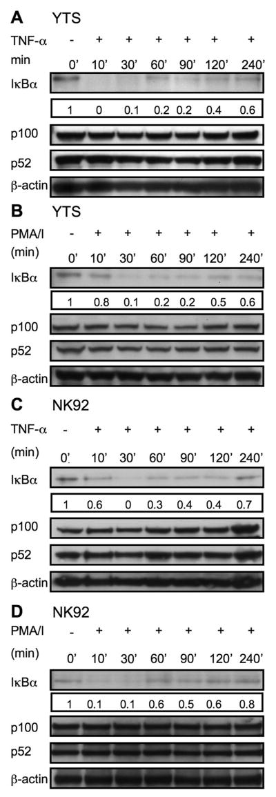
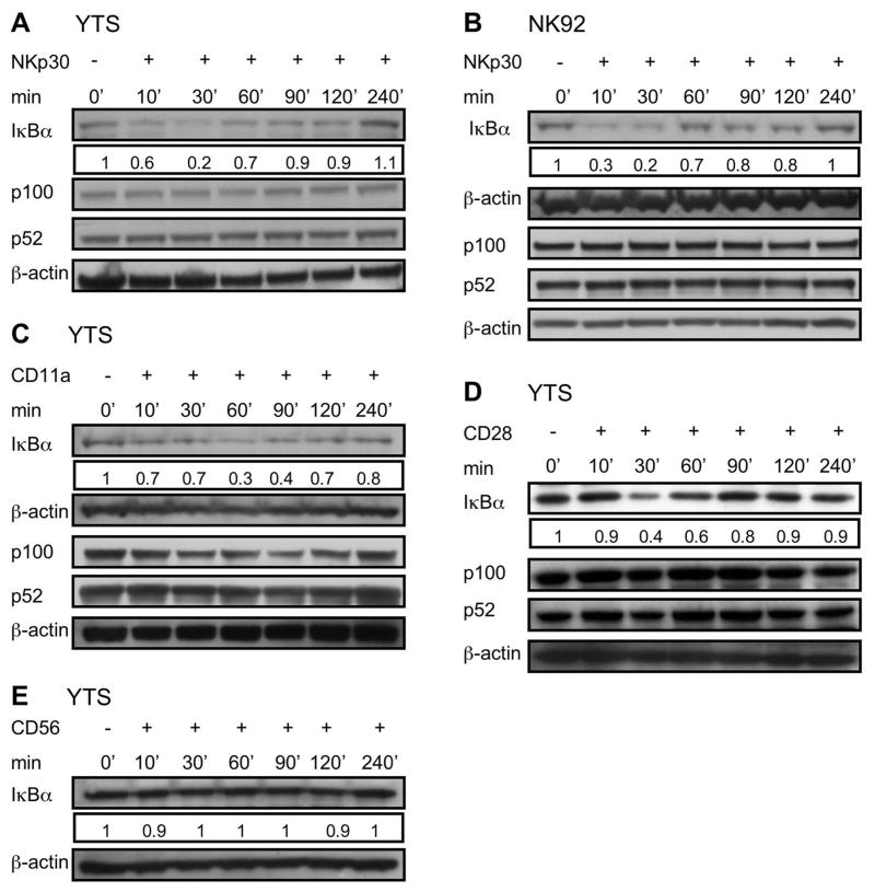
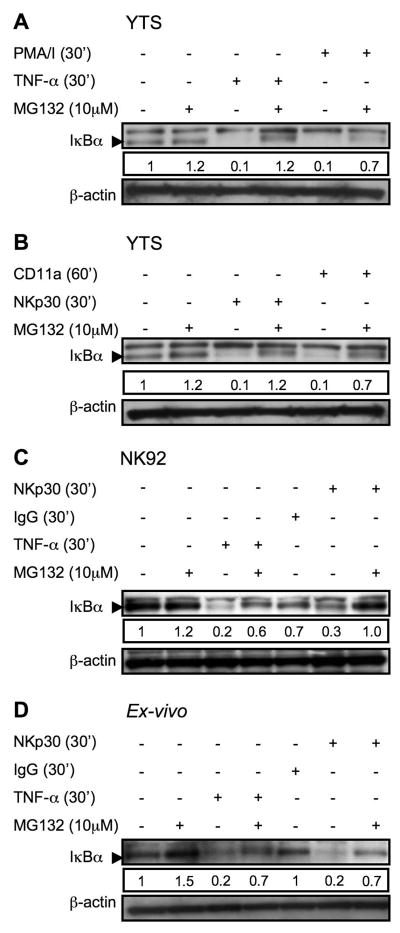
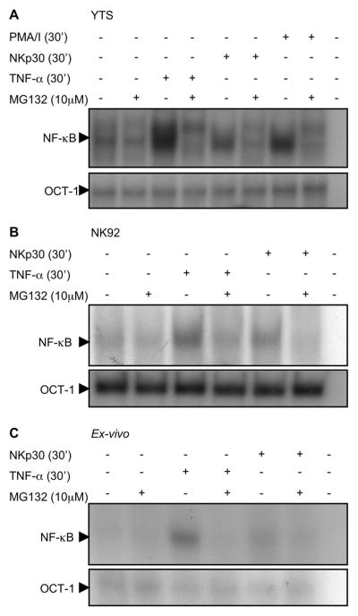
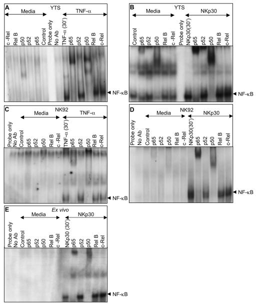
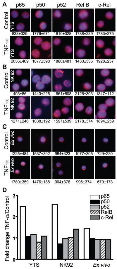
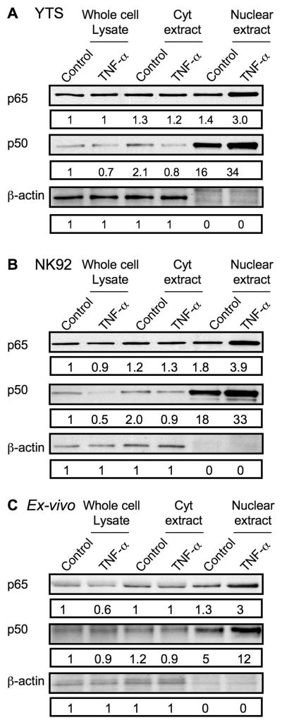
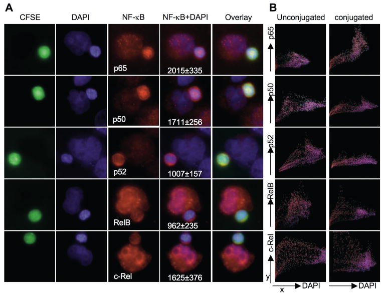
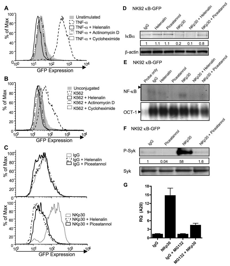
Studies of patients with congenital immunodeficiency due to mutation of the NF-kappaB essential modulator (NEMO) gene have demonstrated that NEMO integrity is required for NK cell cytotoxicity. Thus, we have studied the physiology of NF-kappaB activation in NK cells during the cytolytic program. In resting ex vivo human NK cells or cell lines, IkappaB was degraded after 10 min exposure to PMA and ionomycin, or TNF and was maximally degraded by 30 min. Ligation of several NK cell activation receptors including NKp30 induced a similar response and was blocked by pretreatment with the proteosome inhibitor MG132. There was no short-term effect on p100 processing, the signature of noncanonical NF-kappaB activation. NK cell IkappaB degradation corresponded to increases in nuclear NF-kappaB as detected by EMSA. Supershift of stimulated NK cells and fluorescence microscopy of individual NK cells in cytolytic conjugates demonstrated that the p65/p50 heterodimer was the primary NF-kappaB used. NF-kappaB function was evaluated in NK92 cells transduced with a kappaB GFP reporter, and their conjugation with K562 cells or ligation of NKp30 ligation resulted in rapid GFP accumulation. The latter was prevented by the Syk inhibitor piceatannol. Thus, NK cell activation signaling specifically induces transcriptional activation and synthesis of new NF-kappaB dependent proteins during the initiation of cytotoxicity.


Conflict of interest statement

Disclosures

The authors have no conflicts of interest.

Figures

Figure 1
Figure 1. Rapid activation signal induced IκBα degradation in YTS and NK92 cells
The ability of TNF-α and PMA/I to induce IκB degradation was determined in YTS (A, B) and NK92 (C, D) cells. Cells were treated in solution for the time indicated, lysed, separated on Nu-Page gels and transferred to membranes which were probed with anti-IκBα pAb (~33KD), stripped and reprobed with anti-β-actin rabbit pAb (~38KD) (as a control for protein loading). Processing of p100 (~100KD) to p52 (~52KD) after TNF-α and PMA/I stimulation for the indicated time points was also assessed in lysates with anti-NF-κBp52 rabbit pAb. Numbers below the IκBα blots represent the ratio of the band intensity to the 0 min band intensity for IκBα. Representative results of three independent experiments are shown.
Figure 2
Figure 2. Activating receptor specific IκBα degradation in YTS and NK92 cells
YTS (A, C, D, E) and NK92 cells (B) were exposed to immobilized anti-NKp30 (A, B), anti-CD11a (C), anti-CD28 (D) and anti-CD56 (E) for 0, 10, 30, 60, 90, 120 and 240 min and then lysed on ice. Lysates were separated, transferred and membranes probed with anti-IκBα rabbit pAb (~33KD), stripped and reprobed with anti-β-actin rabbit pAb (~38KD) (as a control for protein loading). Processing of p100 to p52 after exposure to immobilized antibodies was also assessed for each. Numbers below IκBα blot represents the ratio of the band intensity to the 0 min band intensity for IκBα. Representative results of three independent experiments are shown.
Figure 3
Figure 3. Inhibition of activation induced IκBα degradation in YTS, NK92 and ex-vivo NK cells by MG132
YTS cells were pretreated with MG132 for 30 min, restimulated with TNF-α or PMA/I (A), anti-CD11a or anti-NKp30 (B). NK 92 (C) and ex vivo NK cells (D) were pretreated with MG132 for 30 min, restimulated with TNF or anti-NKp30 for 30 min and then lysed. Lysates were separated, transferred and membranes probed with anti-IκBα rabbit pAb (~33KD), as demonstrated by the arrowhead. Membranes were then stripped and reprobed with anti-βactin rabbit pAb (~38KD) as a control for protein loading. A nonspecific band above the IκBα band was found with some lots of the pAb. Numbers below each IκBα blot represent the ratio of the band intensity to the 30 min band intensity for IκBα. Representative results of three independent experiments are shown.
Figure 4
Figure 4. Increased NF-κB nuclear translocation in NK cells after activation
Nuclear protein extracts of YTS (A), NK92 (B) and ex-vivo NK cells (C) were evaluated for NF-κB (top) and Oct-1 (bottom) DNA binding activity by EMSA. Prior to lysis cells were activated with PMA/I (YTS only) or TNF-α or anti-NKp30. Cells were also pretreated with MG132. Nuclear extracts were preincubated with radiolabelled double stranded oligonucleotide NF-κB or Oct-1 probe and reaction products were analyzed on 5% non-denaturing polyacrylamide gels. Selected sections of the autoradiogram are shown. Representative results of three independent experiments are shown.
Figure 5
Figure 5. Activation induced nuclear translocation of specific NF-κB heterodimers in NK cells
Supershift analysis with antibodies to p65, p52, p50, Rel-B and c-Rel was performed on nuclear protein extracts of YTS (A, B), NK92 (C, D) and ex-vivo (E) NK cells. NK cells were treated with TNF-α (A, C) or anti-NKp30 (B, D, E) for 30 minutes. Nuclear extracts preincubated with NF-κB specific Abs were radiolabelled with double stranded oligonucleotide NF-κB probe and reaction products were analyzed on 5% non-denaturing polyacrylamide gels. Selected sections of the autoradiogram are shown highlighting the region of NF-κB bound probe and supershifted bands. Representative results of three independent experiments are shown.
Figure 6
Figure 6. Single NK cell NF-κB nuclear translocation after activation
Untreated (top) and TNF-α treated (bottom) YTS (A), NK92 (B) and ex-vivo (C) NK cells were fixed and stained with DAPI (blue) as well as anti-p65, anti-p50, anti-p52, anti-RelB or anti-c-Rel followed by AlexaFluor 568-conjugated secondary Ab (red). Cells were imaged using fluorescence microscopy to show a merged overlay of DAPI and different NF-κB family members. The numbers beneath each image represent the mean nuclear MFI ± SD from ≥50 cells measured using Volocity. (D) The ratio of mean fluorescence intensity in the nucleus to the whole cell was calculated using Volocity and is represented as fold change after TNF-α activation for each NF-κB family member. Each bar represents individual analysis of 50–60 cells.
Figure 7
Figure 7. Cytoplasmic and nuclear levels of NF-κB p65 and p50 protein in resting and activated NK cells
(A) YTS, (B) NK92, and (C) ex vivo NK cells were treated with control media or TNF-α for 30 minutes and equal amounts of protein from whole cell lysates (left), cytoplasmic extracts (Cyt, middle), and nuclear extracts (right) were evaluated for the presence of NF-κB p65 (top), NF-κB p50 (middle), and β-actin by Western blot. Numbers below each band represent the ratio of the band intensity to that of the band for the whole cell lysate of control-treated cells.
Figure 8
Figure 8. NF-κB nuclear translocation in single NK cells after physiologic activation
Ex vivo NK cells were stained with CFSE and conjugated with K562 cells. (A) Conjugates were fixed and stained with DAPI (blue) and anti-p65, anti-p50, anti-p52, anti-RelB or anti-c-Rel followed with Alexa Fluor 568-conjugated secondary Ab. Fluorescence for CFSE (green), DAPI (blue), NF-κB family members (red), an overlay of NF-κB and DAPI and merged overlay fluorescent intensities are shown. Representative conjugated cells are shown. Mean nuclear MFI ± SD of NF-κB family members in ≥30 ex vivo NK cells conjugated to K562 target cells are shown beneath the NF-κB+DAPI overlay image. (B) Colocalization of individual pixel fluorescent intensities within a single unconjugated NK cell (left) or NK cell conjugated to K562 target cell (right). The x-axis represents DAPI fluorescence intensity and y-axis NF-κB family member fluorescence intensity. Pixels with no DAPI fluorescence localize to the cytoplasm. Plots are representative of 10 analyses.
Figure 9
Figure 9. Activation-induced NF-κB function in NK92 cells
NK92 cells transduced with a κB-GFP reporter construct were stimulated with TNF-α (A), K562 target cells (B), or immobilized IgG (top) or anti-NKp30 (bottom) (C) for 8h after 30 min pretreatment with media, helenalin, actinomycin-D, cycloheximide or piceatannol. GFP expression in live cells was analyzed by FACS and representative results of five independent experiments are shown. NK92 κB-GFP cells were pretreated for 30min with media, helenalin, or piceatannol and stimulated for 30min with immobilized IgG or anti-NKp30. Whole cell lysates were evaluated for the presence of IκBα by Western blot (D) and nuclear extracts for NF-κB (top) and OCT-1 (bottom) DNA binding activity by EMSA (E). Whole cell lysates of NK92 κB-GFB cells pretreated for 30 min with media or piceatannol and stimulated for 30 min with IgG or NKp30 were evaluated by Western blot for phospho-Syk (F, top). The membrane was stripped and reprobed for total Syk (F, bottom). The numbers below the phospho-Syk blot represent the ratio of the phospho-Syk band intensity to the IgG phospho-Syk band. (G) A20 transcripts were quantified using real-time PCR from NK92-κB GFP cells that were pretreated for 30 min with media, or MG132 and then stimulated for 4 h with immobilized IgG or anti-NKp30.

References

    1. Moretta L, Moretta A. Unravelling natural killer cell function: triggering and inhibitory human NK receptors. Embo J. 2004;23:255–259. - PMC - PubMed
    1. Orange JS, Ballas ZK. Natural killer cells in human health and disease. Clin Immunol. 2006;118:1–10. - PubMed
    1. Pende D, Parolini S, Pessino A, Sivori S, Augugliaro R, Morelli L, Marcenaro E, Accame L, Malaspina A, Biassoni R, Bottino C, Moretta L, Moretta A. Identification and molecular characterization of NKp30, a novel triggering receptor involved in natural cytotoxicity mediated by human natural killer cells. J Exp Med. 1999;190:1505–1516. - PMC - PubMed
    1. Hayden MS, West AP, Ghosh S. NF-κB and the immune response. Oncogene. 2006;25:6758–6780. - PubMed
    1. Scheidereit C. IkappaB kinase complexes: gateways to NF-κB activation and transcription. Oncogene. 2006;25:6685–6705. - PubMed

Publication types

MeSH terms