Comparative Study
. 2007 Nov 27;104(48):18907-12.
doi: 10.1073/pnas.0709326104. Epub 2007 Nov 19.

A role for DNA-mediated charge transport in regulating p53: Oxidation of the DNA-bound protein from a distance

Affiliations
Comparative Study

A role for DNA-mediated charge transport in regulating p53: Oxidation of the DNA-bound protein from a distance

Katherine E Augustyn et al. Proc Natl Acad Sci U S A. .

Abstract

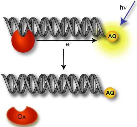
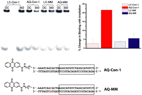
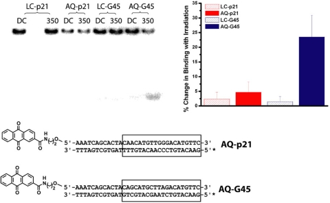
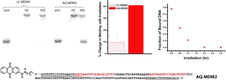
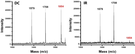
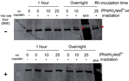
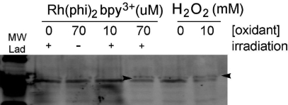
Charge transport (CT) through the DNA base pairs provides a means to promote redox reactions at a remote site and potentially to effect signaling between molecules bound to DNA. Here we describe the oxidation of a cell-cycle regulatory protein, p53, from a distance through DNA-mediated CT. A consensus p53 binding site as well as three DNA promoters regulated by p53 were synthesized containing a tethered DNA photooxidant, anthraquinone. Photoinduced oxidation of the protein occurs from a distance; introduction of an intervening CA mismatch, which inhibits DNA-mediated CT, prevents oxidation of p53. DNA-mediated oxidation is shown to promote dissociation of p53 from only some promoters, and this sequence-selectivity in oxidative dissociation correlates with the biological regulation of p53. Under severe oxidative stress, effected here through oxidation at long range, p53 dissociates from a promoter that activates DNA repair as well as the promoter for the negative regulator of p53, Mdm2, but not from a promoter activating cell-cycle arrest. Mass spectrometry results are consistent with disulfide bond formation in p53 upon DNA-mediated oxidation. Furthermore, DNA-bound p53 oxidation is shown in vivo by up-regulation of p53 and subsequent irradiation in the presence of a rhodium photooxidant to give a new p53 adduct that can be reversed with thiol treatment. This DNA-mediated oxidation of p53 parallels that seen by treating cells with hydrogen peroxide. These results indicate a unique mechanism using DNA-mediated CT chemistry by which p53 activity on different promoters may be controlled globally under conditions of oxidative stress.


Conflict of interest statement

The authors declare no conflict of interest.

Figures

Fig. 1.
Fig. 1.
Schematic illustration of DNA-mediated CT to promote oxidation and dissociation of p53 (red), where AQ serves as the tethered, distally bound photooxidant. Upon photoexcitation, the AQ injects an electron hole into the DNA duplex that can migrate through the DNA to p53, resulting in protein oxidation. This oxidation can induce a conformational change in the DNA-binding domain of p53, promoting its dissociation from the DNA.
Fig. 2.
Fig. 2.
Oxidation of p53 bound to the consensus sequence with and without a base-pair mismatch. The electrophoretic mobility-shift assay (Upper Left) is shown as an autoradiogram after electrophoresis under nondenaturing conditions of LC-Con-1, the consensus sequence without appended oxidant, LC-MM, the consensus sequence containing a CA mismatch, AQ-Con-1, the consensus sequence with tethered AQ and AQ-MM, the mismatch-containing consensus sequence with tethered AQ both in the absence of irradiation (DC), and with irradiation for 30 min at 350 nm (350) in the presence of p53. The sequences for AQ-Con-1 and AQ-MM are shown (Lower) with the p53 binding site boxed and the site of 32P end-labeling indicated by an asterisk; the CA mismatch is highlighted in red. A bar graph (Upper Right) quantifying the percentage change in binding with irradiation for the different sequences with and without the AQ photooxidant also is shown. The percentage change in binding was determined as a ratio of the percentage of bound DNA in the dark control to that in the irradiated sample. Error bars reflect the standard deviations obtained from multiple trials. Samples contained 0.125 μM duplex, 0.5 μM p53, 5 μM dAdT, 0.1% np-40, 0.1 mg/ml BSA in 20 mM Tris·Cl (pH 8), 20% glycerol, 100 mM KCl, and 0.2 mM EDTA.
Fig. 3.
Fig. 3.
Sequence-selectivity in photooxidation of p53. Shown (Upper Left) is an autoradiogram for the gel-shift assay after electrophoresis under nondenaturing conditions of LC-p21, AQ-p21, LC-G45, and AQ-G45 both without irradiation (DC) and with irradiation for 30 min at 350 nm (350) in the presence of p53. The sequences of AQ-p21 and AQ-G45 with the p53 binding sites boxed are indicated (Lower), where the asterisk indicates the site of 32-P end-labeling. A bar graph (Upper Right) quantifying the percentage change in binding with irradiation for the different sequences with and without the AQ photooxidant also is shown. The percentage change in binding was determined as a ratio of the percentage of bound DNA in the dark control to that in the irradiated sample. Error bars reflect the standard deviations obtained from multiple trials. Samples contained 0.125 μM duplex, 0.5 μM p53, 5 μM dAdT, 0.1% np-40, 0.1 mg/ml BSA in 20 mM Tris·Cl (pH 8), 20% glycerol, 100 mM KCl, and 0.2 mM EDTA.
Fig. 4.
Fig. 4.
Oxidation of p53 bound to the Mdm2 promoter. The gel-shift assay (Upper Left) shows an autoradiogram after electrophoresis under nondenaturing conditions of LC-MDM2, the sequence without appended oxidant and AQ-MDM2, the sequence containing a tethered AQ both in the absence of irradiation (DC) and with irradiation for 45 min at 350 nm (350) in the presence of p53. Lanes marked −p53 contain only DNA. Quantification of the percentage change in binding also is shown in a bar graph (Upper Center), where the error bars reflect standard deviations from multiple trials. A time course for p53 dissociation from AQ-MDM2 also is shown (Upper Right) as a function of irradiation. The Mdm2 sequence tethered with AQ is indicated (Lower) (the complementary 32P-labeled strand is not shown), where the p53 binding sites are highlighted in red and the primers used are underlined. Samples contained 0.125 μM duplex, 0.5 μM p53, 5 μM dAdT, 0.1% np-40, 0.1 mg/ml BSA in 20 mM Tris·Cl (pH 8), 20% glycerol, 100 mM KCl, and 0.2 mM EDTA.
Fig. 5.
Fig. 5.
MALDI-TOF mass spectrometry of a p53 tryptic digest after incubation with AQ-Con-1 without (DC) or with (IR) 3 h of irradiation at 350 nm. Samples contained 0.3 μM duplex and 1.2 μM p53 in 20 mM Tris·Cl (pH 8), 20% glycerol, 100 mM KCl, and 0.2 mM EDTA. The fragment with a mass of 1,854 corresponds to the fragment TCPVQWVDSTPPPGTR containing cysteine 141.
Fig. 6.
Fig. 6.
Reversible oxidation of p53 from HCT116 cells visualized by Western blotting analysis. After treatment of HCT116 cells with cisplatin to up-regulate p53, cells were incubated with different concentrations of the rhodium photooxidant for either 1 h or overnight and irradiated with a solar simulator. Cellular protein was analyzed by SDS/PAGE followed by Western blotting analysis. Cells irradiated without rhodium show no p53 adduct bands. Overnight incubation of irradiated cells with either 10 or 25 μM [Rh(phi)2bpy]3+ lead to a new, lower-mobility p53 adduct (red arrowhead, upper gel). Addition of 2-mercaptoethanol removes the oxidation-dependent p53 adduct (lower gel).
Fig. 7.
Fig. 7.
Western blotting analysis of oxidized p53 derived from HeLa cells. Cells were incubated with hydrogen peroxide for 30 min at 37°C (no irradiation) or with [Rh(phi)2(bpy)]3+ for 30 min at 37°C and then irradiated with a solar simulator (UV cut-off filter <345 nm). Total protein was analyzed by SDS/PAGE followed by Western blotting with anti-p53 antibody and a fluorescent secondary antibody. Cells irradiated without rhodium, or in the presence of rhodium but without irradiation, show little change in the p53 banding pattern. However, incubation with 70 μM rhodium with irradiation or hydrogen peroxide yields a change in the p53 profile with a new, lower-mobility band as indicated by an arrowhead.

References

    1. Vogelstein B, Lane D, Levine AJ. Nature. 2000;408:307–310. - PubMed
    1. Vousden KH, Lu X. Nat Rev Cancer. 2002;2:594–604. - PubMed
    1. Prives C, Hall PA. J Pathol. 1999;187:112–126. - PubMed
    1. Brooks CL, Gu W. Mol Cell. 2006;21:307–315. - PMC - PubMed
    1. El-Deiry WS, Tokino T, Velculescu VE, Levy DB, Parsons R, Trent JM, Lin D, Mercer WE, Kinzler KW, Vogelstein B. Cell. 1993;75:817–825. - PubMed

Publication types

MeSH terms