. 2008 Jan;4(1):51-8.
doi: 10.1038/nchembio.2007.40. Epub 2007 Nov 18.

Unnatural substrates reveal the importance of 8-oxoguanine for in vivo mismatch repair by MutY

Affiliations

Unnatural substrates reveal the importance of 8-oxoguanine for in vivo mismatch repair by MutY

Alison L Livingston et al. Nat Chem Biol. 2008 Jan.

Abstract

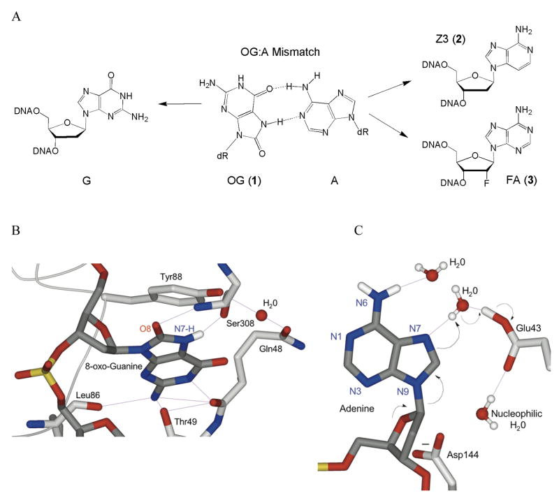
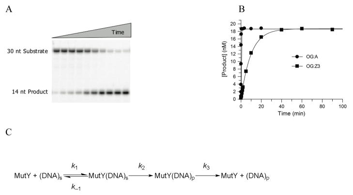
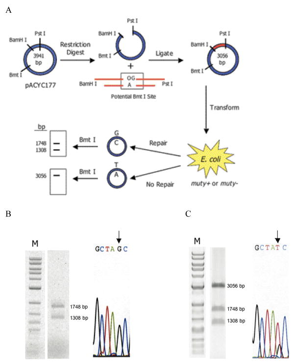
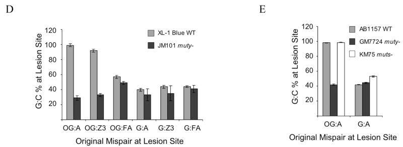
Escherichia coli MutY has an important role in preventing mutations associated with the oxidative lesion 7,8-dihydro-8-oxo-2'-deoxyguanosine (OG) in DNA by excising adenines from OG.A mismatches as the first step of base excision repair. To determine the importance of specific steps in the base pair recognition and base removal process of MutY, we have evaluated the effects of modifications of the OG.A substrate on the kinetics of base removal, mismatch affinity and repair to G-C in an E. coli-based assay. Notably, adenine modification was tolerated in the cellular assay, whereas modification of OG resulted in minimal cellular repair. High affinity for the mismatch and efficient base removal required the presence of OG. Taken together, these results suggest that the presence of OG is a critical feature that is necessary for MutY to locate OG.A mismatches and select the appropriate adenines for excision to initiate repair in vivo before replication.


Figures

Figure 1
Figure 1. Recognition of an OG:A mismatch by D144N BsMY and modifications made to probe features of mismatch repair
(a) Structure of an OG:A base pair and the substitutions made of the mismatch that were examined (b and c) Close-up views of recognition of OG (b) and A (c) from the X-ray crystal structure of D144N Bacillus stearothermophilus MutY bound to an OG:A mismatch-containing duplex. In both panels, portions of the BsMY backbone and important residues are shown in light gray, while DNA is rendered in dark gray. Atoms are colored red (oxygen), blue (nitrogen), and gold (phosphorus), while implied protons are white. Hydrogen bonding interactions are shown as purple lines. Panel B highlights the first step of the proposed dissociative mechanism of MutY, with black arrows representing the flow of electrons. Asn144 is represented as the natural Asp144 to emphasize its proposed role in stabilizing an oxocarbenium ion intermediate. Images generated from pdb file 1RRQ from the World Wide Protein Data Bank, based on data from ref. .
Figure 2
Figure 2. Kinetic Analysis of the Glycosylase Activity of MutY
(a) A representative storage phosphor autoradiogram of the PAGE analysis of strand scission at Z3 (creating 14 nt fragment) upon treatment of the OG:Z3-containing duplex (duplex 2) with MutY at 30 mM NaCl, followed by NaOH quenching. (b) Plotting of the data from panel A, with comparison to that obtained with an OG:A-containing duplex 2 under the same conditions. The data are fitted to a single-exponential to determine the rate constant, k2. (c) Minimal kinetic scheme used for analysis of MutY glycosylase activity
Figure 3
Figure 3. Cell-based MutY-mediated repair assay
(a) Schematic representation of the assay. A mismatch-containing oligonucleotide is ligated into a plasmid vector and transformed into E. coli (mutY+ or mutY−). Repair or correct replication of the mismatch generates a G:C bp that creates a second Bmt I restriction site. The amount of G:C bp is determined via restriction fragment analysis of the recovered plasmid. The amount of repair mediated by MutY is determined by the % G:C bp created in cells containing MutY relative to those lacking MutY. (b and c) Restriction fragment and sequence analysis of lesion-containing plasmids rescued from E. coli. Representative data depicting initial plasmid containing an OG:A mismatch that has been completely converted to G:C (b). and initial plasmid containing a G:A mismatch that has not been converted completely to G:C (c). Gels shown are EtBr-stained agarose (M = 1 kb DNA ladder). Sequencing portion shown is of the BmtI recognition sequence and the arrow points to the location of the lesion. Levels of repair (percent G:C at lesion site) mediated by MutY are assessed by quantification of the agarose gels in which the percent G:C represents an average of at least three separate experiments in which > 100 colonies were pooled and amplified for the digestion analysis. (d and e) Summary graph of percent G:C bp determined from restriction fragment analysis of various base pair mismatches in E coli strains XL-1 Blue and JM101 mutY (d) and AB1157, GM7724, and KM75 (e). Repair is related to the amount of G:C base pairs produced at the lesion site determined by BmtI restriction fragment analysis of mismatch-containing plasmid isolated from E. coli possessing MutY versus lacking MutY. Data represent mean values and error bars represent standard deviation of the sample set, which include at least three separate experiments.

Similar articles

Cited by

References

    1. Iyer RR, Pluciennik A, Burdett V, Modrich PL. DNA Mismatch Repair: Functions and Mechanisms. Chem Rev. 2006;106:302–323. - PubMed
    1. Su SS, Lahue RS, Au KG, Modrich P. Mispair Specificity of Methyl-directed DNA Mismatch Correction. J Biol Chem. 1988;263:6829–6835. - PubMed
    1. Lu AL, Chang DY. Repair of Single Base-Pair Transversion Mismatches of Escherichia coli in Vitro: Correction of Certain A/G Mismatches is Independent of dam methylation and Host mut HLS gene Functions. Genetics. 1988;118:593–600. - PMC - PubMed
    1. Radicella JP, Clark EA, Fox MS. Some mismatch repair activities in Escherichia coli. Proc Natl Acad Sci USA. 1988;85:9674–9678. - PMC - PubMed
    1. Ngheim Y, Cabrera M, Cupples CG, Miller JH. The mutY gene: A mutator locus in Escherichia coli that generates G:C to T:A transversions. Proc Natl Acad Sci USA. 1988;85:2709–2713. - PMC - PubMed

Publication types

MeSH terms

LinkOut - more resources