. 2008 Jan;123(1):129-38.
doi: 10.1111/j.1365-2567.2007.02737.x. Epub 2007 Nov 20.

Neuropilin-1 expression identifies a subset of regulatory T cells in human lymph nodes that is modulated by preoperative chemoradiation therapy in cervical cancer

Affiliations

Neuropilin-1 expression identifies a subset of regulatory T cells in human lymph nodes that is modulated by preoperative chemoradiation therapy in cervical cancer

Alessandra Battaglia et al. Immunology. 2008 Jan.

Abstract

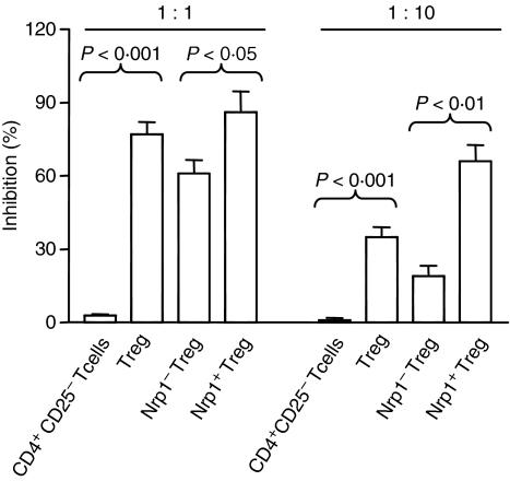
We examined the phenotype and function of CD4+ T cells expressing the semaphorin III receptor neuropilin-1 (Nrp1) in human lymph nodes and peripheral blood. In lymph nodes, Nrp1 identified a small regulatory CD4+ CD25(high) T-cell subpopulation (Nrp1+ Treg) that expressed higher levels of Forkhead box P3 (Foxp3) message and protein than Nrp1- Treg, and various molecular markers of activated Treg, i.e. CD45RO, human leucocyte antigen (HLA)-DR and glucocorticoid-induced tumour necrosis factor receptor (GITR). Similarly to conventional Treg, Nrp1+ Treg proliferated poorly in vitro, and exerted contact-dependent in vitro suppression of T-cell proliferation and cytokine secretion. However, Nrp1+ Treg were more efficient than Nrp1- Treg at inducing suppression. Nrp1 was also expressed on a small subpopulation of CD25(int) and CD25- CD4+ T cells that expressed more Foxp3, CD45RO, HLA-DR and GITR than their Nrp1- counterparts. In contrast, in peripheral blood Nrp1 identified a minor CD4+ T-cell subset that did not display the phenotypic features of Treg lacking Foxp3 expression and marginally expressing CD25. Hence, the function of Nrp1+ CD4+ T cells seemingly depends on their anatomical location. In a previous report, we proposed that Treg may curb the anti-tumour T-cell response in cervical cancer. We show here that Treg and Nrp1+ Treg levels dropped in the tumour-draining lymph nodes of patients with cervical cancer following preoperative chemoradiotherapy in a direct relationship with the reduction of tumour mass, suggesting that suppressor cell elimination facilitated the generation of T cells mediating the destruction of the neoplastic cells left behind after cytotoxic therapy.


Figures

Figure 1
Figure 1
Nrp1 expression on CD4+ T cells in human LN and PB. (a) Nrp1 expression in relation to CD25 staining intensity in CD4+ T cells in LN (top plot) and PB (bottom plot). Most Nrp1+ cells in LN are CD25high whereas in PB Nrp1 expression is essentially restricted to CD25 cells. (b) Gating of Nrp1+ T cells in LN (top left plot) and PB (bottom left plot) and assessment of their Foxp3 and CD25 expression in LN (top right plot) and PB (bottom right plot). Quadrants are drawn based on fluorescence signal generated by samples stained with isotype-matched control antibody for PC5-CD25 and an eightfold molar excess of unlabelled anti-Foxp3 mAb followed by FITC-conjugated anti-Foxp3 mAb for FITC-Foxp3. Numbers in plots represent the percentage of cells in each region.
Figure 2
Figure 2
Responsiveness to polyclonal stimulation of unfractionated Treg, Nrp1+ Treg, and Nrp1 Treg from LN. The proliferative responsiveness to polyclonal stimulation of the unfractionated Treg population and Treg subset defined by Nrp1 coexpression (all 5 × 10⁴ cells/well) isolated with marker-specific antibodies in combination with immunomagnetic beads as described in Materials and methods was compared to that of control CD4+ CD25 T cells. Cells were aseptically loaded with CFSE as described in Materials and methods and the proliferative response (proliferation index) was assessed on day 5 by modfit™/Cell Proliferation Model™ software. Mean values (± SD) obtained in four separate experiments are reported. Significance was assessed by anova followed by Dunnet’s multiple comparison test.
Figure 3
Figure 3
Suppressive activity of unfractionated Treg, Nrp1+ Treg, and Nrp1 Treg from LN on mitogen-driven PBL proliferation. The functional activity of immunomagnetically purified Nrp1+ Treg, Nrp1 Treg, unfractionated Treg population and CD4+ CD25 T cells as a control was tested in a proliferation assay using CFSE-labelled autologous responder PBL immunomagnetically depleted of CD25-expressing cells. Responder cells (5 × 10⁴/well) were cultured in the presence of suppressor cells at 1 : 1 and 1 : 10 suppressor : responder ratio. The proliferative response was assessed on day 5 by computing CFSE halving by modfit™/Cell Proliferation Model™ software. Mean values (±SD) obtained in five separate experiments are reported. Significance was assessed by Student’s t-test.
Figure 4
Figure 4
Unfractionated Treg and Treg subsets defined by Nrp1 expression from LN suppress the secretion of IL-2 and IFN-γ by cocultured CD25-depleted autologous PBL. Culture supernatants from the same cultures described in Fig. 3 were collected at day 2 and cytokines were determined by multiplex ELISA. Data are expressed as per cent variation of cytokine secretion of CD25-depleted autologous PBL (5 × 10⁴/well). Mean values (±SD) obtained in three separate experiments are reported.
Figure 5
Figure 5
Nrp1 is not required for the suppressive activity of Nrp1+ Treg. CFSE-labelled autologous responder CD25-depleted PBL were cocultured with Nrp1+ Treg in the presence of 20 μg/ml anti-Nrp1 M01 mAb or mouse immunoglobulin as control. Alternatively, Nrp1+ Treg were first preincubated with anti-Nrp1 BDCA4 mAb, washed and mixed with CFSE-labelled autologous responder CD25-depleted PBL. The proliferative response was assessed on day 5 by computing CFSE halving by modfit™/Cell Proliferation Model™ software. Mean values (±SD) obtained in two separate experiments are reported.
Figure 6
Figure 6
Foxp3 message in unfractionated Treg, Nrp1+ Treg, and Nrp1 Treg from LN. RNA from unfractionated Treg and Treg subsets defined by Nrp1 expression was prepared as described in Materials and methods. CD4+ CD25 T cells served as control. The cDNA was tested for Foxp3 expression by real-time RT-PCR using CD3ε mRNA expression as reference for normalization. Measurements were performed in triplicates and results are presented as Foxp3 expression relative to CD3ε. Representative data (averages and SD) from three separate experiments are shown.
Figure 7
Figure 7
Treg and Nrp1+ Treg in TDLN are modulated by preoperative chemoradiation therapy in CC patients in direct relationship with pathological response. TDLN-derived cells were stained with FITC anti-CD4, PE anti-Nrp1, ECD anti-CD3, and PE-Cy5 anti-CD25 mAbs. (a) CR did not modify the percentage of Treg within CD4+ T cells. (b) CR decreased Nrp1+ cells within Treg, although not significantly. Following CR, a significant reduction of Treg and Nrp1+ cells within Treg was seen in patients with a complete/near complete disappearance of the primary tumour mass (PR1) compared to patients with a macroscopic residual tumour (PR2) (insets in a and b, respectively). Mean values (±SD).

References

    1. Zwar TD, Van Driel IR, Gleeson PA. Guarding the immune system: suppression of autoimmunity by CD4CD25 immunoregulatory T cells. Immunol Cell Biol. 2006;84:487–501. - PubMed
    1. Dejaco C, Duftner C, Grubeck-Loebenstein B, Schirmer M. Imbalance of regulatory T cells in human autoimmune diseases. Immunology. 2006;117:289–300. - PMC - PubMed
    1. Zou W. Regulatory T cells, tumour immunity and immunotherapy. Nat Rev Immunol. 2006;6:295–307. - PubMed
    1. Baecher-Allan C, Brown JA, Freeman GJ, Hafler DA. CD4+ CD25high regulatory cells in human peripheral blood. J Immunol. 2001;167:1245–53. - PubMed
    1. Baecher-Allan C, Wolf E, Hafler DA. MHC class II expression identifies functionally distinct human regulatory T cells. J Immunol. 2006;176:4622–31. - PubMed

MeSH terms