. 2007 Nov 20:8:49.
doi: 10.1186/1471-2121-8-49.

Regulation of tight junction assembly and epithelial morphogenesis by the heat shock protein Apg-2

Affiliations

Regulation of tight junction assembly and epithelial morphogenesis by the heat shock protein Apg-2

Saima Aijaz et al. BMC Cell Biol. .

Abstract

Background: Tight junctions are required for epithelial barrier formation and participate in the regulation of signalling mechanisms that control proliferation and differentiation. ZO-1 is a tight junction-associated adaptor protein that regulates gene expression, junction assembly and epithelial morphogenesis. We have previously demonstrated that the heat shock protein Apg-2 binds ZO-1 and thereby regulates its role in cell proliferation. Here, we addressed the question whether Apg-2 is also important for junction formation and epithelial morphogenesis.

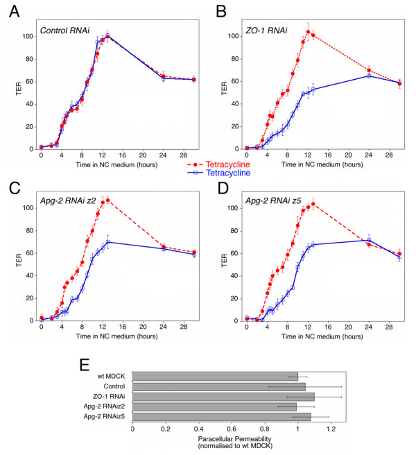
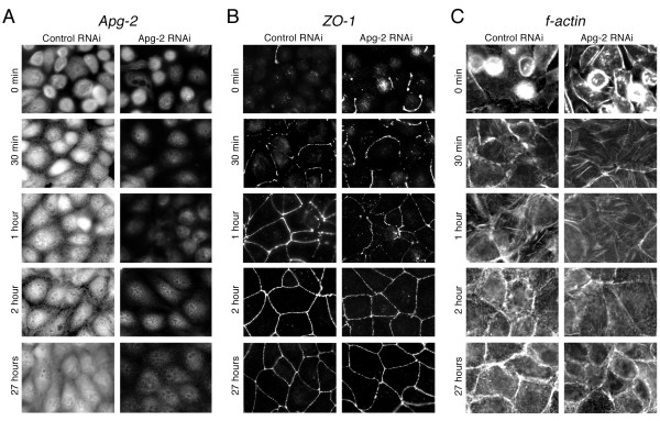
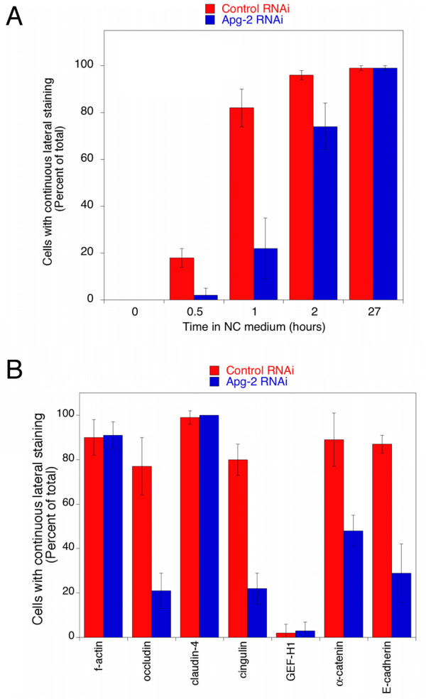
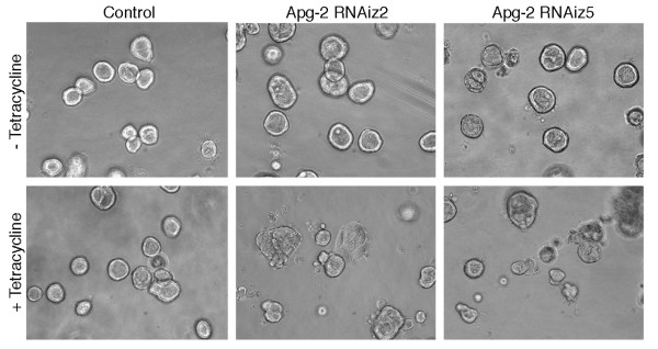
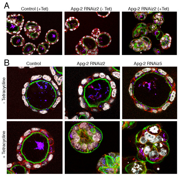
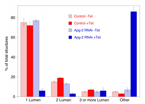
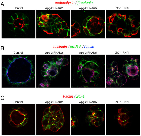
Results: We demonstrate that depletion of Apg-2 by RNAi in MDCK cells did not prevent formation of functional tight junctions. Similar to ZO-1, however, reduced expression of Apg-2 retarded de novo junction assembly if analysed in a Ca-switch model. Formation of functional junctions, as monitored by measuring transepithelial electrical resistance, and recruitment of tight and adherens junction markers were retarded. If cultured in three dimensional extracellular matrix gels, Apg-2 depleted cells, as previously shown for ZO-1 depleted cells, did not form hollow polarised cysts but poorly organised, irregular structures.

Conclusion: Our data indicate that Apg-2 regulates junction assembly and is required for normal epithelial morphogenesis in a three-dimensional culture system, suggesting that Apg-2 is an important regulator of epithelial differentiation. As the observed phenotypes are similar to those previously described for ZO-1 depleted cells and depletion of Apg-2 retards junctional recruitment of ZO-1, regulation of ZO-1 is likely to be an important functional role for Apg-2 during epithelial differentiation.


Figures

Figure 1
Figure 1
Repression of Apg-2 and ZO-1 by regulated RNA interference. Stable MDCK cell lines expressing the tetracycline repressor as well as shRNAs targeting ZO-1 or Apg-2 (z2 and z5 target different sequences), or a non-targeting shRNA under the control of a tetracycline-regulated promoter were plated in low calcium medium onto permeable culture inserts in the presence or absence of tetracycline. After 18 hours, the cells were switched to normal calcium medium for 24 hours adding tetracycline as indicated. The cells were then lysed in SDS-PAGE sample buffer and analysed by immunoblotting. Shown are immunoblots for Apg-2, ZO-1, ZO-2, ZO-3 and α-tubulin. Expression of ZO-1 was reduced to 24 +/- 7% and of Apg-2 to 22 +/- 6% of the respective levels in control cultures (n = 4).
Figure 2
Figure 2
Development of functional tight junctions. (A-D) Cells were plated and incubated without or with tetracycline as described in figure 1. After switching from low calcium to normal calcium medium, TER was measured at the indicated time points. Shown is a typical experiment using a control RNAi cell line (A), a ZO-1 RNAi cell line (B), and two Apg-2 RNAi cell lines (C and D). Values represent means +/- 1 SD of quadruplicates. (E) Paracellular permeability of fluorescent dextran was measured in the same cultures as those shown in panels A to D 3 days after the initiation of junction formation. Shown are the values obtained with 4 kD FITC dextran with tetracycline-treated cultures. Values represent means +/- 1 SD of quadruplicates. Diffusion of 70 kD dextran was also not affected by depletion of Apg-2 and ZO-1 (not shown).
Figure 3
Figure 3
Retardation of ZO-1 recruitment to forming junctions by Apg-2 depletion. Control and Apg-2 RNAi cell lines were cultured in low calcium with tetracycline and junction formation was then induced for the indicated periods of time before fixation. The cells were then labelled with antibodies against Apg-2 (A), ZO-1 (B), or fluorescent phalloidin (C). If tetracycline was not added and Apg-2 was not depleted, Apg-2 RNAi cell lines formed junctions at the same speed as control RNAi cell lines (not shown). Note, recruitment of ZO-1 to the forming junctional complex was retarded, but the distribution of ZO-1 appeared to be normal at later time points.
Figure 4
Figure 4
Quantification of junction formation. Control and Apg-2 RNAi cells were cultured and processed as described in figure 3. Junction formation was allowed to proceed for the indicated amount of time (A) or for one hour (B). The cells were then stained for either ZO-1 (A) or f-actin, occludin, claudin-4, cingulin, GEF-H1, α-catenin or E-cadherin (B). The fractions of cells expressing the labelled markers along the entire lateral cell membrane were then determined by counting. At least five different fields derived from two independent experiments were counted for each marker and time point. Note, as a large fraction of claudin-4 is continuously at the plasma membrane even before the addition of calcium, its early presence at the plasma membrane does not indicate tight junction formation.
Figure 5
Figure 5
Retardation of junction formation by Apg-2 depletion. Control and Apg-2 RNAi cell lines were cultured and processed as in figure 3 and then stained with antibodies against the transmembrane proteins occludin (A) and claudin-4 (B), the junctional plaque components cingulin (C) and GEF-H1 (D), and the adherens junction proteins α-catenin (E) and E-cadherin (F). Shown are images derived from samples fixed after 1 hour and 27 hours of junction formation. Note, retardation of junctional recruitment was observed for occludin, cingulin, α-catenin and E-cadherin. GEF-H1 became only concentrated at tight junctions after longer time points of junction formation. Claudin-4 was present at the plasma membrane at all time points but only appeared to be concentrated at junctions at later time points. GEF-H1 and claudin-4 distributions were not affected by depletion of Apg-2.
Figure 6
Figure 6
Apg-2 depletion inhibits the formation of regular cysts in 3-D cultures. Control and Apg-2 RNAi cell lines were cultured without or with tetracycline in collagen/matrigel gels. The cells were regularly inspected by phase contrast microscopy to monitor cyst and lumen formation. The shown images were taken 7 days after the cultures had been started. Note, most cysts formed by Apg-2 depleted cells had an irregular shape and lacked a lumen.
Figure 7
Figure 7
Apg-2 depletion inhibits epithelial morphogenesis in 3-D cultures. Control and Apg-2 RNAi cell lines were cultured as in figure 6. After fixation, the samples were labelled with FITC-phalloidin and antibodies against β-catenin (shown in red), the Golgi marker GP73 (shown in blue), and for DNA (shown in white). Panel B shows confocal sections taken at a higher magnification than those in panel A. Note, cysts formed by Apg-2 depleted cells were often not normally polarised and had irregular shapes.
Figure 8
Figure 8
Quantification of 3-D morphogenesis assays. Control and Apg-2 RNAi cell lines were cultured and processed as in figure 6. In two experiments performed with different batches of matrigel, pictures from ten different areas were taken and quantified by counting the number and types of cultures. The formed structures were divided into the following classes: spherical structures with a single lumen; 2 lumen and 3 (or more lumen) but with a normal polarised organisation; and all other structures including cysts with no lumen.
Figure 9
Figure 9
Effect of Apg-2 and ZO-1 depletion on the organisation of 3-D cultures. Control, and Apg-2 and ZO-1 RNAi cell lines were cultured as in figure 6. After fixation, the samples were labelled with antibodies against β-catenin and podocalyxin/GP135 (A); occludin, erbB-2 and with Alexa654-phalloidin (B); or with TRITC-phalloidin and anti-ZO-1 antibodies (C). Note, erbB-2 does not only localise to the lateral plasma membrane but also to cytoplasmic organelles, causing the cytoplasmic staining between the nucleus in the apical membrane in control cultures (B). Residual ZO-1 staining in ZO-1 RNAi cells was detected at cell-cell contacts (C).

References

    1. Anderson JM, Van Itallie CM, Fanning AS. Setting up a selective barrier at the apical junction complex. Curr Opin Cell Biol. 2004;16:140–145. doi: 10.1016/j.ceb.2004.01.005. - DOI - PubMed
    1. Schneeberger EE, Lynch RD. The tight junction: a multifunctional complex. Am J Physiol. 2004;286:C1213–28. doi: 10.1152/ajpcell.00558.2003. - DOI - PubMed
    1. Cereijido M, Contreras RG, Shoshani L. Cell adhesion, polarity, and epithelia in the dawn of metazoans. Physiol Rev. 2004;84:1229–1262. doi: 10.1152/physrev.00001.2004. - DOI - PubMed
    1. Tsukita S, Furuse M, Itoh M. Multifunctional strands in tight junctions. Nat Rev Mol Cell Biol. 2001;2:286–293. doi: 10.1038/35067088. - DOI - PubMed
    1. Aijaz S, Balda MS, Matter K. Tight Junctions: Molecular Architecture and Function. Int Rev Cytol. 2006;248:261–298. - PubMed

Publication types