. 2007 Nov 21;2(11):e1225.
doi: 10.1371/journal.pone.0001225.

SNP array karyotyping allows for the detection of uniparental disomy and cryptic chromosomal abnormalities in MDS/MPD-U and MPD

Affiliations

SNP array karyotyping allows for the detection of uniparental disomy and cryptic chromosomal abnormalities in MDS/MPD-U and MPD

Lukasz P Gondek et al. PLoS One. .

Abstract

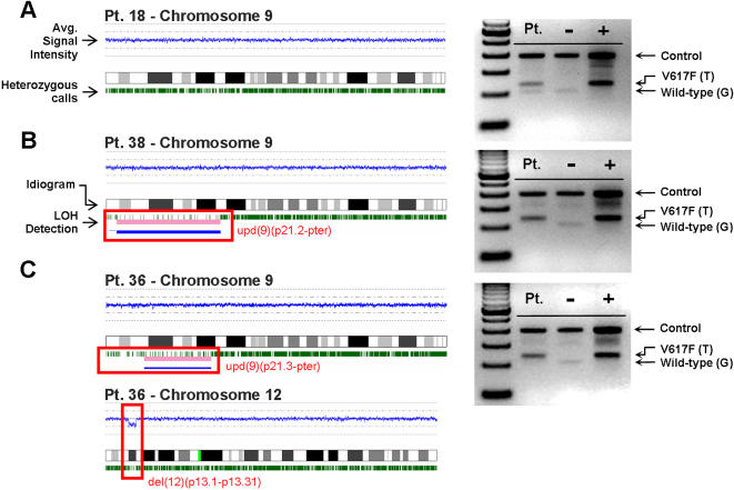
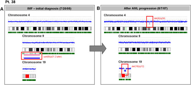
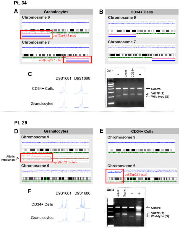
We applied single nucleotide polymorphism arrays (SNP-A) to study karyotypic abnormalities in patients with atypical myeloproliferative syndromes (MPD), including myeloproliferative/myelodysplastic syndrome overlap both positive and negative for the JAK2 V617F mutation and secondary acute myeloid leukemia (AML). In typical MPD cases (N = 8), which served as a control group, those with a homozygous V617F mutation showed clear uniparental disomy (UPD) of 9p using SNP-A. Consistent with possible genomic instability, in 19/30 MDS/MPD-U patients, we found additional lesions not identified by metaphase cytogenetics. In addition to UPD9p, we also have detected UPD affecting other chromosomes, including 1 (2/30), 11 (4/30), 12 (1/30) and 22 (1/30). Transformation to AML was observed in 8/30 patients. In 5 V617F+ patients who progressed to AML, we show that SNP-A can allow for the detection of two modes of transformation: leukemic blasts evolving from either a wild-type jak2 precursor carrying other acquired chromosomal defects, or from a V617F+ mutant progenitor characterized by UPD9p. SNP-A-based detection of cryptic lesions in MDS/MPD-U may help explain the clinical heterogeneity of this disorder.


Conflict of interest statement

Competing Interests: The authors have declared that no competing interests exist.

Figures

Figure 1
Figure 1. SNP karyograms confirm loss of heterozygosity in patients homozygous for JAK2 V617F.
SNP-A based karyotypic analysis on chromosome 9 for (A) a patient heterozygous for the JAK2 V617F mutation and (B,C) two patients homozygous for the JAK2 V617F mutation. (A,B and C, left portion) Signal intensity and SNP karyograms for each corresponding patient; the blue line represents the average fluorescent signal intensity of each SNP and oscillates around the diploid marker line; green tics represent heterozygous calls for each individual SNP. Areas of UPD are associated with the absence of heterozygous calls and are highlighted by blue and pink bars. Extraneous calls in regions of UPD occur as a result of contamination by non-clonal cells. UPD was confirmed by microsatellite analysis (data not shown). (C) In addition to chromosome 9, patient #36 also exhibited a segmental deletion in chromosome 12 as indicated by decreases in the copy number and frequency of heterozygous calls. (A,B, and C, right portion) Corresponding ARMS-PCR analysis of the JAK2 V617F mutation in each patient confirms heterozygous (A) and homozygous (B,C) mutational status (gel images are cropped and enhanced).
Figure 2
Figure 2. SNP-A can be used to identify lesions acquired during AML evolution.
SNP-A karyograms demonstrate that before transformation (A), patient #38 showed only UPD9p at initial diagnosis as a sole abnormality (consistent with a homozygous JAK2 V617F mutation) along with normal chromosomes 4 and 19. However, after transformation to AML (B), repeated SNP-A analysis showed the presence of a V617F- leukemic clone with a normal chromosome 9 and newly-acquired micro-deletions on both chromosomes 4 and 19.
Figure 3
Figure 3. Karyotypic analysis of AML evolution in patients with the JAK2 V617F mutation.
Karyograms illustrate the contributing lesions in patient #34 (A, B) and patient #29 (D, E). In panels C and F, microsatellite analysis confirms the UPD for patients #34 and #29, respectively. In patient #34 (A,B,C), only one distinct clone was identified: in both granulocytes (A) and blasts (B), loss of heterozygosity calls on chromosome 9 confirms UPD9p, consistent with the homozygous JAK2 V617F mutation. In addition, both cell types also possess UPD on 7q. (C, left portion) Microsatellite analysis of blasts and granulocytes confirms LOH on 9p in both cell types. (C, right portion) Electrophoresis gel of ARMS-PCR in patient #34 also shows a homozygous JAK2 V617F mutation in both granulocytes and blasts (gel image is cropped and enhanced). In patient #29 (D,E,F), two distinct clones were identified using DNA isolated from both granulocytes and CD34+ blasts. In granulocytes (D), UPD9p is consistent with a homozygous JAK2 V617F mutation while analysis of chromosome 6 did not reveal any abnormalities. LOH on chromosome 9 was not completely resolved in the karyogram obtained; however, comparative analysis of granulocytes and lymphocytes confirmed allelic imbalance (red line: lymphocytes, green line: granulocytes). In contrast, when CD34- selected blasts were analyzed (E), UPD9p was not identified while a segmental deletion on chromosome 6 was found. In panel F, microsatellite analysis and electrophoresis gel of ARMS-PCR demonstrate the presence of homozygosity for the JAK2 V617F mutation in granulocytes but heterozygosity in CD34+ cells.

References

    1. Andersen CL, Wiuf C, Kruhoffer M, Korsgaard M, Laurberg S, et al. Frequent occurrence of uniparental disomy in colorectal cancer. Carcinogenesis. 2007;28:38–48. - PubMed
    1. Raghavan M, Lillington DM, Skoulakis S, Debernardi S, Chaplin T, et al. Genome-wide single nucleotide polymorphism analysis reveals frequent partial uniparental disomy due to somatic recombination in acute myeloid leukemias. Cancer Res. 2005;65(2):375–378. - PubMed
    1. Lindblad-Toh K, Tanenbaum DM, Daly MJ, Winchester E, Lui WO, et al. Loss-of-heterozygosity analysis of small-cell lung carcinomas using single-nucleotide polymorphism arrays. Nat.Biotechnol. 2000;18(9):1001–1005. - PubMed
    1. Morison IM, Ellis LM, Teague LR, Reeve AE. Preferential loss of maternal 9p alleles in childhood acute lymphoblastic leukemia. Blood. 2002;99(1):375–377. - PubMed
    1. Pei J, Kruger WD, Testa JR. High-resolution analysis of 9p loss in human cancer cells using single nucleotide polymorphism-based mapping arrays. Cancer Genet.Cytogenet. 2006;170(1):65–68. - PubMed

Publication types