. 2007 Dec 19;150(4):915-25.
doi: 10.1016/j.neuroscience.2007.09.061. Epub 2007 Oct 5.

Lasting changes in a network of interneurons after synapse regeneration and delayed recovery of sensitization

Affiliations

Lasting changes in a network of interneurons after synapse regeneration and delayed recovery of sensitization

A K Urazaev et al. Neuroscience. .

Abstract

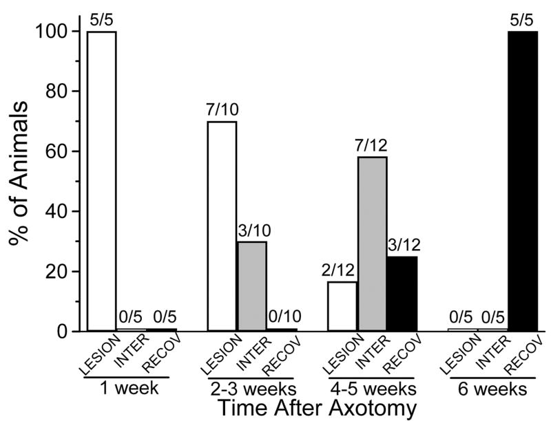
Regeneration of neuronal circuits cannot be successful without restoration of full function, including recovery of behavioral plasticity, which we have found is delayed after regeneration of specific synapses. Experiments were designed to measure neuronal changes that may underlie recovery of function. Sensitization of the leech withdrawal reflex is a non-associative form of learning that depends on the S-interneuron. Cutting an S-cell axon in Faivre's nerve disrupted the capacity for sensitization. The S-cell axon regenerated its electrical synapse with its homologous cell after 3-4 weeks, but the capacity for sensitization was delayed for an additional 2-3 weeks. In the present experiments another form of non-associative conditioning, dishabituation, was also eliminated by S-cell axotomy; it returned following regeneration. Semi-intact preparations were made for behavioral studies, and chains of ganglia with some skin were used for intracellular recording and skin stimulation. In both preparations there was a similar time-course, during 6 weeks, of a lesion-induced decrease and delayed restoration of both S-cell action potential threshold to depolarizing pulses and S-cell firing in response to test stimuli. However, the ability of sensitizing stimuli to decrease S-cell threshold and enhance S-cell activity in response to test stimuli did not fully return after regeneration, indicating that there were lasting changes in the circuit extending beyond the period necessary for full recovery of behavior. Intracellular recordings from the axotomized S-cell revealed a shift in the usual balance of excitatory and inhibitory input, with inhibition enhanced. These results indicate that loss of behavioral plasticity of reflexive shortening following axotomy in the S-cell chain may be related to reduced S-cell activity, and that additional processes underlie full recovery of sensitization of the whole body shortening reflex.


Figures

Figure 1
Figure 1
Schematic diagrams of the preparations. A. Preparation used for behavioral experiments consisted of the head and initial 11 segments. These were intact and, in response to a test stimulus applied anteriorly, exhibited reflexive shorting as measured with a tension transducer. The stronger, sensitizing stimuli were delivered to silver wires tied to the body wall of segment 10. Activity of the S cell was monitored with a suction electrode applied to the connectives posterior to ganglion 13. B. Ganglion 12 was exposed and a suction electrode attached to the posterior connectives for recording S-cell activity. For intracellular recording from the S-cell and from sensory neurons in ganglion 4, the skin remained attached to one side, to which the test stimulation was delivered.
Figure 2
Figure 2
Intracellular recording to measure conduction along the S-cell chain. Configuration as in Fig. 1B, with intracellular recording from S-cell in ganglion 9 (S9) and stimulating suction electrode 1 on anterior connectives at the time indicated by the artifact at the start of each trace. A. Response to anterior connective stimulation in Sham operated animal, in which threshold for the S-cell axon was low and the impulse rapidly propagated along the chain and into the soma of S9. B. In axotomized (Lesion) animals, action potentials generated by the anterior suction electrode (electrode 1) propagated into S4_(data not shown) but not through the lesion, so that only with stronger stimulation producing EPSPs in S9 was it possible to elicit action potentials. C. In 1 of 13 animals in the Early Regeneration group, from 4 to 5 weeks, there was intermittent through-conduction; at higher threshold, EPSPs activated action potentials, but with a greater delay. Conversely, posterior connective stimulation could directly activate action potentials in S4 (not shown), confirming regeneration of the S9 axon. D. Preparation in which the S9 axon had regenerated. Resting potentials were 42–44 mV in A, C, and D and 54 mV in B. Different artifact amplitudes do not indicate different stimulus strengths.
Figure 3
Figure 3
The amplitudes of shortening responses to test stimuli as a percentage of baseline contraction (indicated by a broken line at 100%), ± S.E.M. Individual responses were averaged in four blocks of 5 consecutive responses (abscissa, Blocks 1 through 4). Leeches in each experimental group experienced the sensitizing stimulus immediately following the tests of baseline responding (indicated by the downward arrows). Leeches in each control group did not receive this sensitizing stimulus, but received an identical stimulus after Block 4 (upward arrows). In Panels A–C, data are presented for the group receiving sensitizing training (filled circles) and the control group (open circles). The Mean Normalized Sensitization (MNS) is presented as filled squares in panels A–C and as various symbols in Panel D, as indicated in the inset.
Figure 4
Figure 4
The amplitudes of reflexive shortening, as a percentage of baseline contraction (indicated by broken line at 100%) ± S.E.M, presented according to condition and time relative to axotomy. Thus Shams were without axotomy, Lesions were those without S-cell axon regeneration, Intermediate were those with functional S-cell axon regeneration but without capacity for sensitization of the whole-body shortening reflex, and Recovery were those with both functional S-cell axon regeneration and capacity for sensitization. The individual shortenings following sensitizing stimulation (delivered after baseline stimulation, as indicated by arrow) were grouped in 4 blocks of 5 consecutive responses (abscissa, Blocks 1 through 4).
Figure 5
Figure 5
Regeneration of the S-cell axon preceded recovery of capacity for sensitization. Animals in each weekly timeframe (1-to-6 weeks following axotomy) were divided according to their behavioral and physiological characteristics—whether conduction was interrupted (LESION) or restored without recovery of sensitization (Intermediate, INTER), or was restored with recovery of capacity for sensitization (Recovery, RECOV). The number of animals in each category (above each bar) was plotted across time from axotomy (1, 2–3, 4–5, or 6 weeks). Chi-square test for independence revealed that the variables in the groups of axotomized animals were significantly associated to the time after axotomy (p<0.0001). Asterisk (*) indicates significant difference from Baseline at p<0.05.
Figure 6
Figure 6
Excitability of the S-cell as measured by the number of action potentials in response to a test stimulus. Although whether the S-cell axon had been cut did not affect the baseline number of action potentials to a test stimulus, the operation and the time for recovery affected the response after the sensitizing stimulus. In Sham animals after a sensitizing stimulus, the number of action potentials produced by a test stimulus increased significantly, whereas in lesioned animals before regeneration (Lesion) the number dropped significantly, which was not true following regeneration. Asterisk indicates significant difference from Baseline at p<0.05 (*) and p<0.01 (**).
Figure 7
Figure 7
Excitability of the S-cell in midbody ganglion 4 (S4) as measured by the magnitude of a 20 ms current pulse required to bring the neuron to threshold. There was no significant difference between baseline thresholds among the groups, although there were differences following the sensitizing stimulus. Whereas in Sham controls the threshold dropped significantly in some time blocks following a sensitizing stimulus, this was not true of the axotomized preparations, including the Late Regen (i.e. late regeneration) preparations.
Figure 8
Figure 8
Responses of S9 to connective stimulation posterior to S12. A, left: In sham-operated animals, the evoked action potentials were direct and not associated with synaptic input. A, middle and right: Following axotomy of S9, an excitatory postsynaptic potential (EPSP) (middle) and an EPSP followed by an inhibitory postsynaptic potential (IPSP) (right) were evoked by stimulation of the posterior connective. B, left: Below threshold for direct activation of the S-cell axon, increasing stimulation sometimes first recruited an EPSP, followed by additional recruitment of an IPSP. B, right: Depolarization of the cell membrane from the resting potential level of −50 mV enhanced the IPSP, while hyperpolarization decreased and abolished it, consistent with an IPSP generated by a conductance increase.
Figure 9
Figure 9
Distribution of sub-threshold synaptic responses in S9 to the stimulation of posterior connective, as in Fig. 8, according to the degree of repair. Posterior connective stimulation did not trigger synaptic activity (open bars), or caused only EPSPs (gray bars), or caused EPSPs followed by IPSPs (black bars) for each experimental group. These data indicated that although the biphasic EPSP/IPSPs were seen in some unoperated preparations, S-cell axotomy considerably increased the likelihood of its appearance in a response to posterior connective stimulation below threshold for the S-cell. χ2-test for independence revealed that the variables in the experimental groups were significantly associated to the incidence of subthreshold synaptic responses to posterior connective stimulation (p<0.0001).

Similar articles

References

    1. Asztalos Z, Baba K, Yamamoto D, Tully T. The fickle mutation of a cytoplasmic tyrosine kinase effects sensitization but not dishabituation in Drosophila melanogaster. J Neurogenet. 2007;21:59–71. - PMC - PubMed
    1. Blinzinger K, Kreutzberg G. Displacement of synaptic terminals from regenerating motoneurons by microglial cells. Z Zellforsch Mikrosk Anat. 1968;85:145–157. - PubMed
    1. Bristol AS, Sutton MA, Carew TJ. Neural circuit of tail-elicited siphon withdrawal in Aplysia. I. Differential lateralization of sensitization and dishabituation. J Neurophysiol. 2004;91:666–677. - PubMed
    1. Burrell BD, Sahley CL, Muller KJ. Non-associative learning and serotonin induce similar bi-directional changes in excitability of a neuron critical for learning in the medicinal leech. J Neurosci. 2001;21:1401–1412. - PMC - PubMed
    1. Burrell BD, Sahley CL, Muller KJ. Differential effects of serotonin enhance activity of an electrically coupled neural network. J Neurophysiol. 2002;87:2889–2895. - PubMed

Publication types

MeSH terms