

Interpreting scan data acquired from multiple scanners: a study with Alzheimer's disease
- PMID: 18032068
- PMCID: PMC2225446
- DOI: 10.1016/j.neuroimage.2007.09.066
Interpreting scan data acquired from multiple scanners: a study with Alzheimer's disease
Abstract
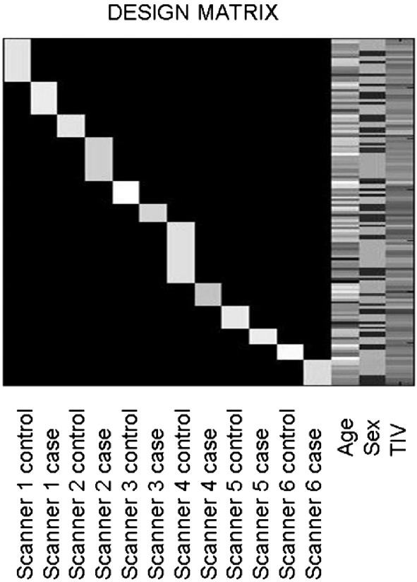
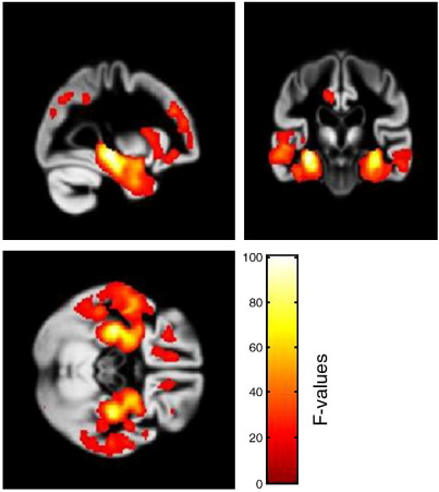
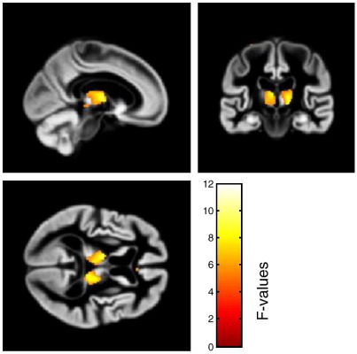
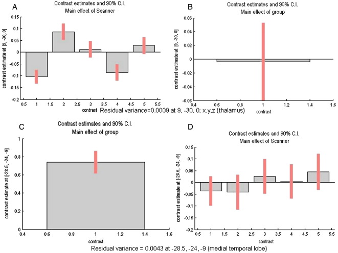
Large, multi-site studies utilizing MRI-derived measures from multiple scanners present an opportunity to advance research by pooling data. On the other hand, it remains unclear whether or not the potential confound introduced by different scanners and upgrades will devalue the integrity of any results. Although there are studies of scanner differences for the purpose of calibration and quality control, the current literature is devoid of studies that describe the analysis of multi-scanner data with regard to the interaction of scanner(s) with effects of interest. We investigated a data-set of 136 subjects, 62 patients with mild to moderate Alzheimer's disease and 74 cognitively normal elderly controls, with MRI scans from one center that were acquired over 10 years with 6 different scanners and multiple upgrades over time. We used a whole-brain voxel-wise analysis to evaluate the effect of scanner, effect of disease, and the interaction of scanner and disease for the 6 different scanners. The effect of disease in patients showed the expected significant reduction of grey matter in the medial temporal lobe. Scanner differences were substantially less than the group differences and only significant in the thalamus. There was no significant interaction of scanner with disease group. We describe the rationale for concluding that our results were not confounded by scanner differences. Similar analyses in other multi-scanner data-sets could be used to justify the pooling of data when needed, such as in studies of rare disorders or in multi-center designs.
Figures
References
-
- Ashburner J. A fast diffeomorphic image registration algorithm. NeuroImage. 2007;38:95–113. - PubMed
-
- Ashburner J., Friston K.J. Unified segmentation. NeuroImage. 2005;26:839–851. - PubMed
-
- Ashburner J., Friston K.J. Voxel-based morphometry—The methods. NeuroImage. 2000;11:805–821. - PubMed
-
- Benjamini Y., Hochberg Y. Controlling the false discovery rate—A practical and powerful approach to multiple testing. J. R. Stat. Soc. 1995;57:289–300.
-
- Briellmann R.S., Syngeniotis A., Jackson G.D. Comparison of hippocampal volumetry at 1.5 Tesla and at 3 Tesla. Epilepsia. 2001;42:1021–1024. - PubMed
Publication types
MeSH terms
Grants and funding
LinkOut - more resources
Full Text Sources
Medical
