

Uracil recognition by replicative DNA polymerases is limited to the archaea, not occurring with bacteria and eukarya
- PMID: 18032433
- PMCID: PMC2241895
- DOI: 10.1093/nar/gkm1023
Uracil recognition by replicative DNA polymerases is limited to the archaea, not occurring with bacteria and eukarya
Abstract
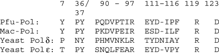
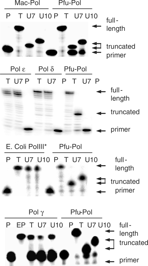
Family B DNA polymerases from archaea such as Pyrococcus furiosus, which live at temperatures approximately 100 degrees C, specifically recognize uracil in DNA templates and stall replication in response to this base. Here it is demonstrated that interaction with uracil is not restricted to hyperthermophilic archaea and that the polymerase from mesophilic Methanosarcina acetivorans shows identical behaviour. The family B DNA polymerases replicate the genomes of archaea, one of the three fundamental domains of life. This publication further shows that the DNA replicating polymerases from the other two domains, bacteria (polymerase III) and eukaryotes (polymerases delta and epsilon for nuclear DNA and polymerase gamma for mitochondrial) are also unable to recognize uracil. Uracil occurs in DNA as a result of deamination of cytosine, either in G:C base-pairs or, more rapidly, in single stranded regions produced, for example, during replication. The resulting G:U mis-pairs/single stranded uracils are promutagenic and, unless repaired, give rise to G:C to A:T transitions in 50% of the progeny. The confinement of uracil recognition to polymerases of the archaeal domain is discussed in terms of the DNA repair pathways necessary for the elimination of uracil.
Figures
References
-
- Fogg MJ, Pearl LH, Connolly BA. Structural basis for uracil recognition by archaeal family B DNA polymerases. Nat. Struct. Biol. 2002;9:922–927. - PubMed
-
- Shuttleworth G, Fogg MJ, Kurpiewski MR, Jen-Jacobson L, Connolly BA. Recognition of the pro-mutagenic base uracil by family B DNA polymerases from archaea. J. Mol. Biol. 2004;337:621–634. - PubMed
-
- Lindahl T. Instability and decay of the primary structure of DNA. Nature. 1993;362:709–715. - PubMed
-
- Pearl LH. Structure and function in the uracil-DNA glycosylase superfamily. Mutat. Res. 2000;460:165–181. - PubMed
Publication types
MeSH terms
Substances
Grants and funding
LinkOut - more resources
Full Text Sources
Other Literature Sources
