. 2008 Feb;82(3):1271-83.
doi: 10.1128/JVI.01405-07. Epub 2007 Nov 21.

Human papillomavirus E1 helicase interacts with the WD repeat protein p80 to promote maintenance of the viral genome in keratinocytes

Affiliations

Human papillomavirus E1 helicase interacts with the WD repeat protein p80 to promote maintenance of the viral genome in keratinocytes

Alexandra Côté-Martin et al. J Virol. 2008 Feb.

Abstract

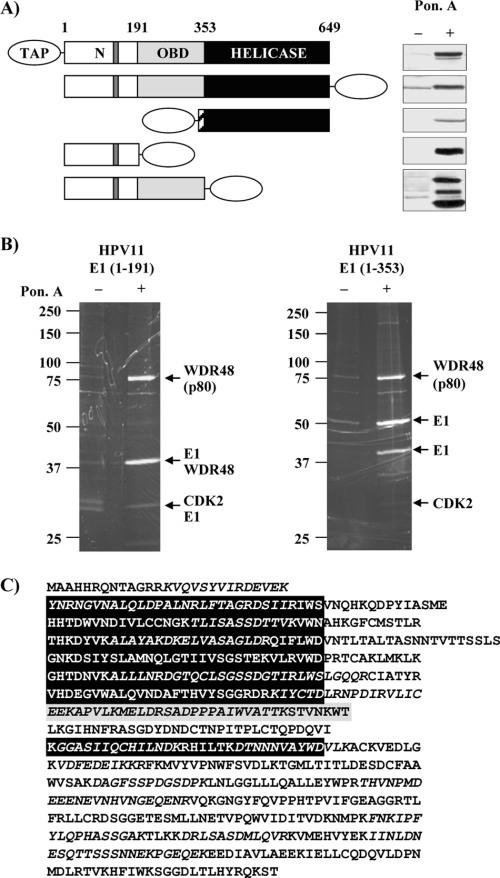
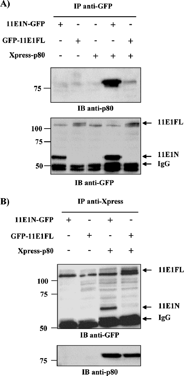
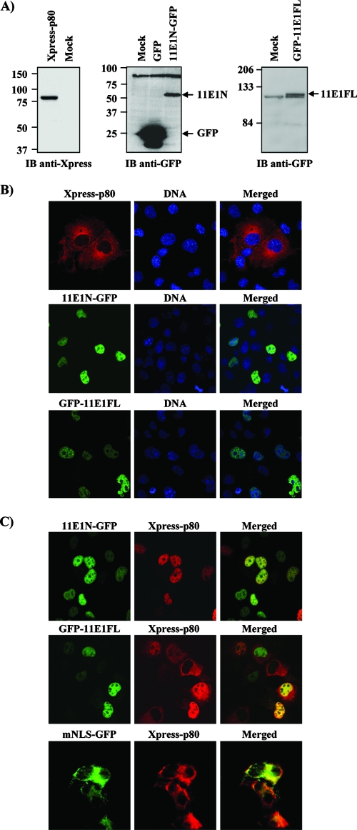
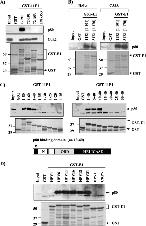
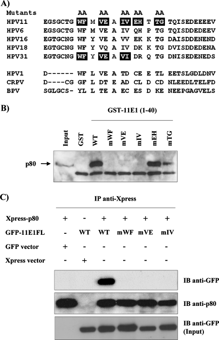
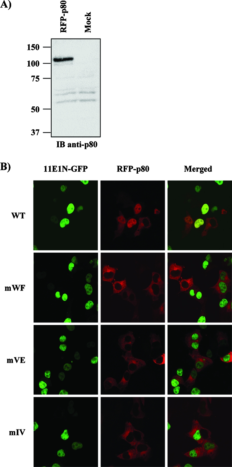
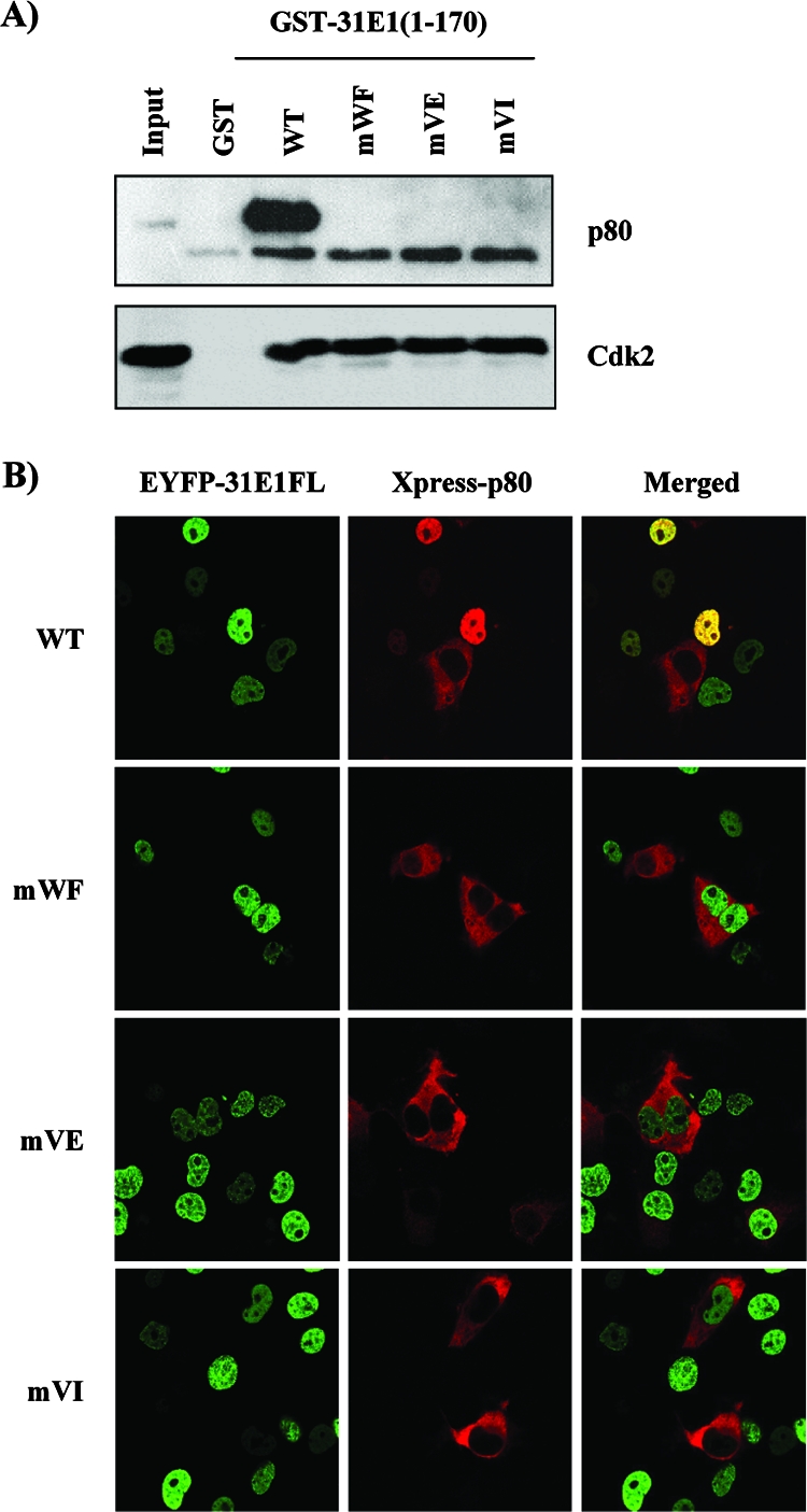
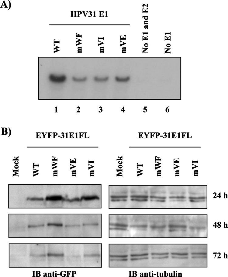
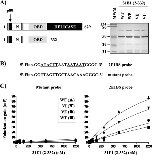
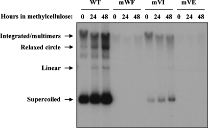
Due to the limited coding capacity of their small genomes, human papillomaviruses (HPV) rely extensively on host factors for the completion of their life cycles. Accordingly, most HPV proteins, including the replicative helicase E1, engage in multiple protein interactions. The fact that conserved regions of E1 have not yet been ascribed a function prompted us to use tandem affinity protein purification (TAP) coupled to mass spectrometry to identify novel targets of this helicase. This method led to the discovery of a novel interaction between the N-terminal 40 amino acids of HPV type 11 (HPV11) E1 and the cellular WD repeat protein p80 (WDR48). We found that interaction with p80 is conserved among E1 proteins from anogenital HPV but not among cutaneous or animal types. Colocalization studies showed that E1 can redistribute p80 from the cytoplasm to the nucleus in a manner that is dependent on the E1 nuclear localization signal. Three amino acid substitutions in E1 proteins from HPV11 and -31 were identified that abrogate binding to p80 and its relocalization to the nucleus. In HPV31 E1, these substitutions reduced but did not completely abolish transient viral DNA replication. HPV31 genomes encoding two of the mutant E1 proteins were not maintained as episomes in immortalized primary keratinocytes, whereas one encoding the third mutant protein was maintained at a very low copy number. These findings suggest that the interaction of E1 with p80 is required for efficient maintenance of the viral episome in undifferentiated keratinocytes.


Figures

FIG. 1.
FIG. 1.
Identification of cellular proteins that interact with HPV11 E1. (A) EcR-293 stable cell lines expressing different TAP-tagged constructs of HPV11 E1 under the control of an ecdysone-inducible promoter. Protein expression was analyzed by Western blotting after a 24-h induction with (+) or without (−) 3 μM ponasterone A (Pon. A), an ecdysone analogue. The structures of the different TAP-tagged E1 proteins are diagrammed on the left. The amino acid boundaries of the N-terminal (N), OBD, and helicase domains are indicated. The location of the CBM is indicated by dark gray bars. The hatched bar in HPV11 E1(353-649) represents the SV40 nuclear localization sequence that was attached to this construct. (B) TAP-purified protein complexes from cells expressing HPV11 E1(1-191) and E1(1-353). Purified proteins were separated by SDS-PAGE and stained with Sypro Ruby. Major protein bands indicated by arrows were excised, trypsin digested, and identified by LC-MS/MS analysis. Some leaky expression of both TAP-tagged constructs led to the purification of E1 and its associated proteins even in uninduced EcR-293 cells, but in much smaller amounts. (C) Amino acid sequence of p80. The seven putative WD domains are aligned and shown in black boxes. A potential eight-WD repeat is highlighted in gray. Tryptic peptides identified by MS are in italics.
FIG. 2.
FIG. 2.
p80 interacts with HPV11 E1 in co-IP assays. co-IP of Xpress-fused p80 with GFP-fused HPV11 E1(1-191) (11E1N) and full-length HPV11 E1(1-649) (11E1FL). HEK293 cells were transfected with the indicated combination of expression vectors and harvested 48 h posttransfection, and WCE were immunoprecipitated by using either anti-GFP (A) or anti-Xpress (B) antibodies. IB, immunoblot; −, absence of; +, presence of.
FIG. 3.
FIG. 3.
Intracellular localization of p80 and HPV11 E1 in COS-7 cells. (A) Expression levels of GFP-E1 and p80 fusion proteins. COS-7 cells were transiently transfected with either 11E1N-GFP, GFP-11E1FL, or Xpress-p80 expression vector. Cells were harvested 24 h posttransfection, and WCE were submitted to Western blot analysis. (B) Cytoplasmic localization of p80 and nuclear localization of 11E1N-GFP and GFP-11E1FL in COS-7 cells. DNA was stained with TO-PRO-3. (C) Cellular colocalization of Xpress-p80 with 11E1N-GFP, GFP-11E1FL, or 11E1NmNLS-GFP in COS-7 cells. IB, immunoblot.
FIG. 4.
FIG. 4.
Mapping of a p80 binding domain on E1 and interaction of p80 with E1 from different PV types. (A) p80 interacts with the N-terminal domain of HPV11 E1. The indicated HPV11 E1 fragments were expressed as GST fusions in bacteria and used in pulldown assays using EcR-293 WCE. Bound proteins were separated by SDS-PAGE and analyzed by Western blotting using antibodies directed against p80 or against Cdk2 as a positive control. The bottom panel shows a Coomassie blue-stained SDS-PAGE gel of the purified GST-E1 proteins that were first immobilized and then eluted from the beads in these pulldown experiments. The sizes of molecular weight markers (in thousands) are indicated on the left. (B) Binding of p80 from HeLa and C33A cells to the N-terminal domains of HPV11 and HPV31 E1. The indicated HPV11 and corresponding HPV31 E1 fragments were expressed as GST fusions and used in pulldown assays using HeLa and C33A WCE, as indicated. Bound proteins were separated by SDS-PAGE and analyzed by Western blotting using antibodies directed against p80. The bottom panel shows an SDS-PAGE gel of the GST-E1 proteins eluted from the beads. (C) Mapping of p80 interaction domain on HPV11 E1. Short N-terminal (N) peptides of HPV11 E1 were expressed as GST fusions in bacteria and tested for p80 binding in pulldown assays as indicated above. The location of the p80 interaction domain on E1 is illustrated. The bottom panel shows an SDS-PAGE gel of the purified GST-E1 proteins eluted from the beads. (D) Interaction of p80 with E1 from different PV types. E1 N-terminal domains of one cutaneous HPV type (1), two genital low-risk HPV (types 6 and 11), three genital high-risk HPV (types 16, 18, and 31), BPV1, and CRPV were expressed as GST fusions and tested for p80 binding in pulldown assays as described above. The bottom panel shows an SDS-PAGE gel of the purified GST-E1 proteins eluted from the beads.
FIG. 5.
FIG. 5.
Amino acid substitutions in HPV11 E1 that abrogate its interaction with p80. (A) The top panel shows amino acid sequence alignment of the p80 interaction domains of E1 from different anogenital HPV types, and the lower panel shows the corresponding region from three cutaneous PV types (HPV1, CRPV, and BPV) which do not bind p80. Boxed amino acids were mutated to alanine. (B) Double-alanine substitutions were introduced into GST-11E1(1-40), and the resulting mutant proteins were used in pulldown assays with EcR-293 WCE. Bound proteins were separated by SDS-PAGE and analyzed by Western blotting using a polyclonal anti-p80 antibody. (C) Alanine mutants in GFP-11E1FL abrogate its interaction with p80 in co-IP assays. Plasmids containing GFP-fused WT or mutant 11E1FL were cotransfected with Xpress-p80 expression vector in HEK293 cells. WCE were prepared 48 h posttransfection and submitted to IP by using an anti-Xpress antibody. Precipitated proteins were analyzed by Western blot analysis using anti-GFP antibody or anti-p80 antibody as an IP control. The lower panel shows the amount of input GFP-E1 proteins present in the WCE prior to IP. IB, immunoblot; −, absence of; +, presence of.
FIG. 6.
FIG. 6.
Mutant E1 proteins fail to relocalize p80 from the cytoplasm to the nucleus. (A) Expression of RFP-p80 fusion protein. COS-7 cells were transiently transfected with the indicated expression vectors and harvested 24 h posttransfection, and WCE were submitted to Western blot analysis with an anti-p80 antibody. (B) COS-7 cells were cotransfected with RFP-p80 and WT or mutant 11E1N-GFP expression vectors for cellular localization studies.
FIG. 7.
FIG. 7.
HPV31 E1 proteins defective for p80 interaction. (A) The three E1 double-amino-acid substitutions previously shown in HPV11 E1 to abrogate interaction with p80 were introduced into a fusion of the HPV31 E1 N-terminal domain to GST. The resulting mutant proteins were purified from bacteria and used in pulldown experiments with EcR-293 WCE. Bound proteins were analyzed by Western blot analysis using a polyclonal anti-p80 antibody. Western blot analysis using antibody against Cdk2 is shown as control. (B) Effect of E1 substitutions on p80 cellular localization. The substitutions were introduced into pEYFP-HPV31 E1 expression vector. WT or mutant EYFP-31E1 plasmids were cotransfected with Xpress-p80 in COS-7 cells, and the cellular localization of the encoded proteins was analyzed by confocal microscopy.
FIG. 8.
FIG. 8.
Transient replication activity of mutant HPV31 E1 proteins. (A) Transient HPV DNA replication. C33A cells were transfected with 3 μg of the WT (lane 1) or the indicated mutant E1 (lanes 2 to 4) expression vector plus 1 μg of the E2 expression vector and 1 μg of the HPV31Luc vector, which contains the ori. As negative controls, cells were transfected with the origin-containing vector alone (no E1 and E2 [lane 5]) or with the E2 and HPV31Luc vectors (no E1 [lane 6]). (B) Accumulation of WT and mutant EYFP-HPV31 E1. The levels of the indicated E1 proteins in transfected C33A cells were detected by Western blotting with an anti-GFP antibody 24, 48, and 72 h posttransfection (left panels). Mock-transfected cells were used as a negative control. Western blot analysis with an anti-β-tubulin antibody (right panels) was performed to ensure that equal amounts of total cellular proteins were loaded in each lane.
FIG. 9.
FIG. 9.
Effect of amino acid substitutions that abrogate the p80 interaction on binding of HPV31 E1 to the origin in vitro. (A) Schematic representations of the full-length HPV31 E1 protein and of the 332-aa-long fragment that was expressed and purified from E. coli either in WT form or containing the WF, VE, or VI substitution. The right panel shows a Coomassie blue-stained gel of the purified fragments (3 μg of each were loaded on the gel). The band migrating at about 70 kDa is likely DnaK. The positions and sizes (in thousands) of molecular weight markers (MWM) are shown on the left. N, N-terminal domain. (B) Nucleotide sequences of the two fluorescein-labeled probes containing, respectively, two E1 binding sites (2E1BS, underlined and bolded) and two mutated sites. Only the sequence of the labeled DNA strand is shown. (C) Fluorescence polarization binding isotherms were determined with 15 nM of the indicated probe and increasing concentrations of the indicated E1 fragment. Each isotherm was determined in triplicate; errors bars (indicating standard deviations) are barely visible on the graph as they are smaller than the symbols. Polarization values were determined as described in Materials and Methods.
FIG. 10.
FIG. 10.
Maintenance and amplification of mutant HPV31 genomes in primary human keratinocytes. Southern blot analysis showing the status of the viral genome in HFK immortalized with either WT HPV31 (WT) or mutant derivatives bearing the indicated E1 amino acid substitutions that abrogate p80 binding. HPV31-immortalized HFK were induced to differentiate in methylcellulose-containing medium for 24 and 48 h, as indicated.

Similar articles

Cited by

References

    1. Amin, A. A., S. Titolo, A. Pelletier, D. Fink, M. G. Cordingley, and J. Archambault. 2000. Identification of domains of the HPV11 E1 protein required for DNA replication in vitro. Virology 272137-150. - PubMed
    1. Bairoch, A., R. Apweiler, C. H. Wu, W. C. Barker, B. Boeckmann, S. Ferro, E. Gasteiger, H. Huang, R. Lopez, M. Magrane, M. J. Martin, D. A. Natale, C. O'Donovan, N. Redaschi, and L. S. Yeh. 2005. The Universal Protein Resource (UniProt). Nucleic Acids Res. 33D154-D159. - PMC - PubMed
    1. Campbell, R. E., O. Tour, A. E. Palmer, P. A. Steinbach, G. S. Baird, D. A. Zacharias, and R. Y. Tsien. 2002. A monomeric red fluorescent protein. Proc. Natl. Acad. Sci. USA 997877-7882. - PMC - PubMed
    1. Chiang, C. M., M. Ustav, A. Stenlund, T. F. Ho, T. R. Broker, and L. T. Chow. 1992. Viral E1 and E2 proteins support replication of homologous and heterologous papillomaviral origins. Proc. Natl. Acad. Sci. USA 895799-5803. - PMC - PubMed
    1. Clower, R. V., J. C. Fisk, and T. Melendy. 2006. Papillomavirus E1 protein binds to and stimulates human topoisomerase I. J. Virol. 801584-1587. - PMC - PubMed

Publication types

MeSH terms

LinkOut - more resources