Clinical Trial
. 2008 Feb;82(3):1448-57.
doi: 10.1128/JVI.01409-07. Epub 2007 Nov 21.

Phase I trial of a CD8+ T-cell peptide epitope-based vaccine for infectious mononucleosis

Affiliations
Clinical Trial

Phase I trial of a CD8+ T-cell peptide epitope-based vaccine for infectious mononucleosis

Suzanne L Elliott et al. J Virol. 2008 Feb.

Abstract

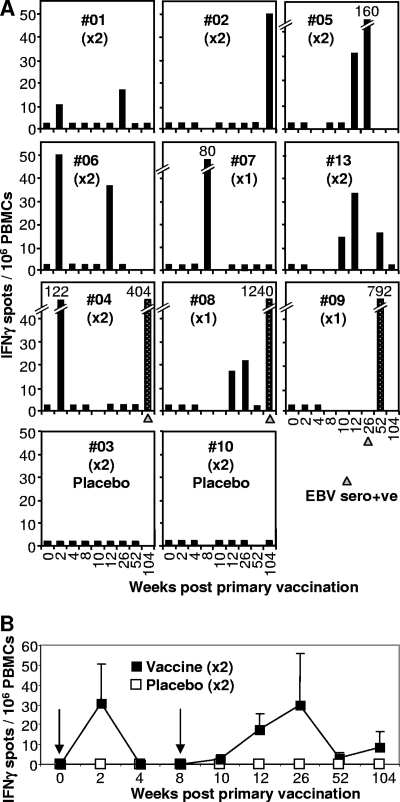
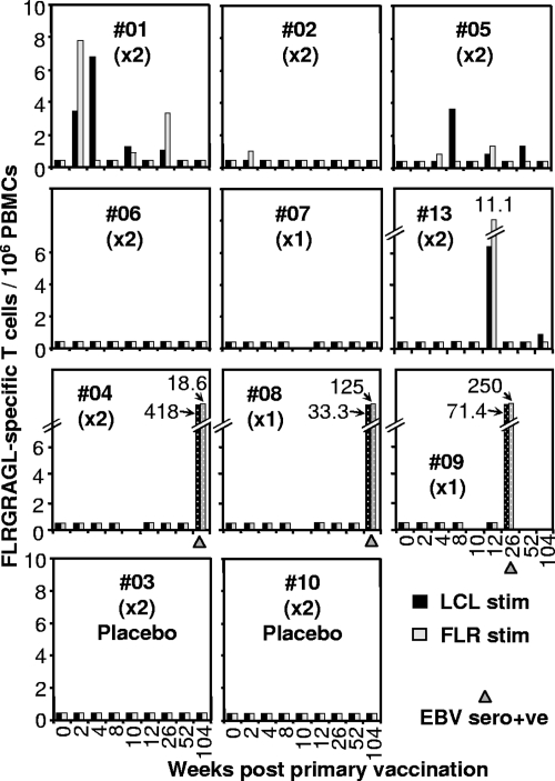
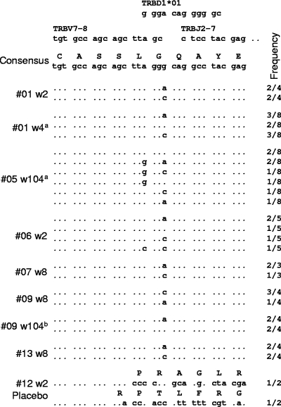
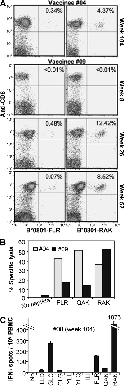
A single blind, randomized, placebo-controlled, single-center phase I clinical trial of a CD8(+) T-cell peptide epitope vaccine against infectious mononucleosis was conducted with 14 HLA B*0801-positive, Epstein-Barr virus (EBV)-seronegative adults. The vaccine comprised the HLA B*0801-restricted peptide epitope FLRGRAYGL and tetanus toxoid formulated in a water-in-oil adjuvant, Montanide ISA 720. FLRGRAYGL-specific responses were detected in 8/9 peptide-vaccine recipients and 0/4 placebo vaccine recipients by gamma interferon enzyme-linked immunospot assay and/or limiting-dilution analysis. The same T-cell receptor Vbeta CDR3 sequence that is found in FLRGRAYGL-specific T cells from most EBV-seropositive individuals could also be detected in the peripheral blood of vaccine recipients. The vaccine was well tolerated, with the main side effect being mild to moderate injection site reactions. After a 2- to 12-year follow-up, 1/2 placebo vaccinees who acquired EBV developed infectious mononucleosis, whereas 4/4 vaccinees who acquired EBV after completing peptide vaccination seroconverted asymptomatically. Single-epitope vaccination did not predispose individuals to disease, nor did it significantly influence development of a normal repertoire of EBV-specific CD8(+) T-cell responses following seroconversion.


Figures

FIG. 1.
FIG. 1.
Ex vivo IFN-γ ELISPOT analysis. (A) The vaccine was administered twice at week 0 and week 8 (×2), except for vaccinees #07, #08, and #09, who received only the first vaccination (×1). Vaccinees received a 5-μg dose of peptide, except #13, who received a 50-μg dose, and #03 and #10, who received no peptide (placebo). PBMC from vaccinees collected prior to vaccination (week 0) and at the indicated number of weeks after the first vaccination were analyzed by ex vivo IFN-γ ELISPOT assay using the FLR peptide and frozen PBMC. Three vaccinees seroconverted asymptomatically within 2 years (#04, #08, and #09), and the time at which serology first indicated EBV seroconversion is indicated with a gray triangle. Vaccine-induced responses are represented by black bars. Speckled bars illustrate responses after EBV seroconversion. Black bars at 2.5 IFN-γ spots/106 PBMC represent no significant response and are shown to indicate that an assay was performed on PBMC collected at that time point. (B) Mean numbers of IFN-γ spots (± standard errors) for vaccinees #01, #02, #04, #05, #06, and #13 (black squares), who received vaccinations at 0 and 8 weeks (arrows). The mean numbers of spots for all four placebo recipients are also shown (white squares). The two groups are significantly different (P = 0.041) by analysis of variance, which included a term for weeks.
FIG. 2.
FIG. 2.
LDA. Fresh PBMC from the same vaccines as described in the legend to Fig. 1 were analyzed by LDA. Cultures were stimulated with either autologous LCLs (black bars) or FLR peptide (10 μg/ml) (gray bars) and assessed in 51Cr release assays. Speckled bars illustrate responses after EBV seroconversion. The time when serology first indicated EBV seroconversion is indicated with a gray triangle. Bars at 0.4/106 PBMC represent no significant response and are shown to indicate that an assay was performed on PBMC collected at that time point.
FIG. 3.
FIG. 3.
TCR Vβ sequences postvaccination. Sequences from cloned PCR products from PBMC or bulk cultures from vaccine recipients taken at the indicated times postvaccination (w, week) are shown. The frequency with which the indicated sequence was found over the total number of clones providing TCR sequence is indicated in the right column. a, from bulk cultures; b, vaccinee #09 had seroconverted at this time. The consensus sequence for LC13 (3) is shown at the top together with the germ line gene sequences that are used in the generation of this CDR3 region. Clear PCR products were obtained from #01 PBMC collected at weeks 2, 10, 26, 68, and 104; from #01 bulk cultures at week 4; from #05 bulk cultures at week 104; from #06 PBMC at weeks 2, 12, and 26; from #06 PBMC at weeks 2, 12, and 26; from #07 PBMC at week 8; from #09 PBMC at weeks 8, 10, 12, and 52; from placebo #12 PBMC at week 2; and from #13 PBMC at weeks 8 and 10. No detectable PCR products were obtained from #01 PBMC at week 0 (two samples), placebo #10 PBMC at weeks 2 and 4, placebo #11 PBMC at weeks 2 and 4, and placebo #12 PBMC at week 12 (data not shown).
FIG. 4.
FIG. 4.
CD8 T-cell response of vaccinees after EBV seroconversion. (A) PBMC from vaccinees (no. 9 and 4) collected at the indicated time points postvaccination were assessed by FACS using FLR-specific (left panels, B*0801-FLR) and RAK-specific (right panels, B*0801-RAK) pentamers (x axis) and anti-CD8 monoclonal antibody (y axis). The percentage of CD8 T cells that are pentamer positive are indicated in the top right corner of each panel. (B) Standard chromium release assay using bulk cultures generated from PBMC from vaccinees #09 (week 52) and #04 (week 104) to asses lytic activity specific for FLR, QAK, and RAK. (C) Ex vivo IFN-γ ELISPOT assay using PBMC from vaccinee #08 (collected at week 104) to asses T-cell responses to the HLA A*0201-restricted epitopes LLDFVRFMGV (LLD), GLCTLVAML (GLC), CLGGLLTMV (CLG), ILIYNGWYA (ILI), YLLEMLWRL (YLL), and YLQQNWWTL (YLQ) and the HLA B*0801-restricted epitopes FLR, QAK, and RAK.

References

    1. Adams, L. A., B. Deboer, G. Jeffrey, R. Marley, and G. Garas. 2006. Ganciclovir and the treatment of Epstein-Barr virus hepatitis. J. Gastroenterol. Hepatol. 211758-1760. - PubMed
    1. Aichele, P., K. Brduscha-Riem, S. Oehen, B. Odermatt, R. M. Zinkernagel, H. Hengartner, and H. Pircher. 1997. Peptide antigen treatment of naive and virus-immune mice: antigen-specific tolerance versus immunopathology. Immunity 6519-529. - PubMed
    1. Argaet, V. P., C. W. Schmidt, S. R. Burrows, S. L. Silins, M. G. Kurilla, D. L. Doolan, A. Suhrbier, D. J. Moss, E. Kieff, T. B. Sculley, and I. S. Misko. 1994. Dominant selection of an invariant T cell antigen receptor in response to persistent infection by Epstein-Barr virus. J. Exp. Med. 1802335-2340. - PMC - PubMed
    1. Bharadwaj, M., S. R. Burrows, J. M. Burrows, D. J. Moss, M. Catalina, and R. Khanna. 2001. Longitudinal dynamics of antigen-specific CD8+ cytotoxic T lymphocytes following primary Epstein-Barr virus infection. Blood 982588-2589. - PubMed
    1. Bharadwaj, M., M. Sherritt, R. Khanna, and D. J. Moss. 2001. Contrasting Epstein-Barr virus-specific cytotoxic T cell responses to HLA A2-restricted epitopes in humans and HLA transgenic mice: implications for vaccine design. Vaccine 193769-3777. - PubMed

Publication types

MeSH terms