. 2008 Feb;82(3):1547-57.
doi: 10.1128/JVI.01976-07. Epub 2007 Nov 21.

Induction of particle polymorphism by cucumber necrosis virus coat protein mutants in vivo

Affiliations

Induction of particle polymorphism by cucumber necrosis virus coat protein mutants in vivo

Kishore Kakani et al. J Virol. 2008 Feb.

Abstract

The Cucumber necrosis virus (CNV) particle is a T=3 icosahedron consisting of 180 identical coat protein (CP) subunits. Plants infected with wild-type CNV accumulate a high number of T=3 particles, but other particle forms have not been observed. Particle polymorphism in several T=3 icosahedral viruses has been observed in vitro following the removal of an extended N-terminal region of the CP subunit. In the case of CNV, we have recently described the structure of T=1 particles that accumulate in planta during infection by a CNV mutant (R1+2) in which a large portion of the N-terminal RNA binding domain (R-domain) has been deleted. In this report we further describe properties of this mutant and other CP mutants that produce polymorphic particles. The T=1 particles produced by R1+2 mutants were found to encapsidate a 1.9-kb RNA species as well as smaller RNA species that are similar to previously described CNV defective interfering RNAs. Other R-domain mutants were found to encapsidate a range of specifically sized less-than-full-length CNV RNAs. Mutation of a conserved proline residue in the arm domain near its junction with the shell domain also influenced T=1 particle formation. The proportion of polymorphic particles increased when the mutation was incorporated into R-domain deletion mutants. Our results suggest that both the R-domain and the arm play important roles in the formation of T=3 particles. In addition, the encapsidation of specific CNV RNA species by individual mutants indicates that the R-domain plays a role in the nature of CNV RNA encapsidated in particles.


Figures

FIG. 1.
FIG. 1.
(A)Tertiary structure of the CNV CP C subunit with ordered arm, S-, and P-domains, with the hinge (h) indicated. The structure is based on homology modeling using the X-ray crystal structure of the TBSV CP subunit (27). The location of the conserved Pro85 residue is also shown. The disordered R-domain is not shown. (B) The CP linear structure is shown, using the same color scheme as in panel A. The number of amino acids in each domain is given. (C) Alignment of the arm region and the junction at the S-domain of several members of the Tombusviridae family, demonstrating conservation of the Pro residue (Pro85 in CNV) (asterisk) in this region. CNV, TBSV, and Cymbidium ringspot virus (CyRSV) are tombusviruses, TCV and Melon necrotic spot virus (MNSV) are carmoviruses, Red clover necrotic mosaic virus (RCNMV) is a dianthovirus, and CLSV (Cucumber leaf spot virus) and PoLV (Pothos latent virus) are aureusviruses.
FIG. 2.
FIG. 2.
Summary of R-domain and arm mutant structures. The top of the diagram shows the 58-aa sequence of the WT CNV CP R-domain along with the downstream 34-aa arm region (open box) and the beginning of the S-domain. Shaded regions in the R-domain correspond to previously identified sequences found to be important for particle accumulation (unpublished data). The C-terminal LRR is indicated above the R-domain sequence. R and arm region mutants are shown beneath. Sequences retained in each mutant are shown. The presence of a proline (P) or glycine (G) at aa position 85 in the arm region of the various mutants is indicated in the shaded box.
FIG. 3.
FIG. 3.
Analysis of R1+2 particles and particle RNA. (A) TEM of particles extracted from leaf tissue infected with the indicated mutant. Particles were stained with 2% uranyl acetate and photographed at a magnification of ×80,000. The bars correspond to approximately 34 nm. (B) Agarose gel of total particles from plants infected with WT CNV (lane 1) and R1+2 infections (lane 2). Lanes 3 and 4 contain the faster- and slower-migrating species, respectively, of sucrose gradient-purified R 1+2 particles. The gels were stained with EtBr or Coomassie blue as indicated. The positions of the two major virion species are shown with arrows. (C) Agarose gel of RNA extracted from total R1+2 particles (lane 2) or sucrose gradient-fractioned particles as in panel B. The numbers on the right correspond to the approximate sizes of the major RNA species.
FIG. 4.
FIG. 4.
Characterization of CNV DI-like RNAs in T=1 particles of mutant R1+2. (A) EtBr-stained agarose gel showing RT-PCR products of RNA purified from T=1 particles of R1+2 (lane 2). RT-PCR primers corresponded to the 5′ and 3′ termini of CNV gRNA. Lane 1 contains a molecular size standard (1-kb Plus DNA ladder; Invitrogen). The approximate sizes of the RT-PCR products are indicated. (B) Structure of R1+2 DI-like RNAs. The structure of CNV genome RNA is shown at the top along with the structures of two previously described prototype CNV DI RNAs (11). The three major regions of the CNV genome that are retained in the prototype DI RNAs (I, II, and III) are depicted by the boxed regions in CNV DI-15 and DI-42. Region III of DI-42 is divided into subregions IIIa and IIIb as previously described (11). The bottom shows structures of DI-like RNAs deduced from nucleotide sequence analysis. The shading patterns used correspond to those of the prototype DI-like RNAs. Numbers at the 5′ and 3′ termini of the boxes indicate the nucleotide positions at the boundaries of the box region relative to CNV gRNA. The size in nucleotides of each DI or DI-like RNA is shown on the right. D-RNA, defective RNA.
FIG. 5.
FIG. 5.
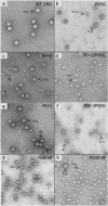
TEM of particles extracted from leaves infected with the indicated mutants. Particles were stained with 2% uranyl acetate and photographed at a magnification of ×80,000. The T=3 or T=1 morphology of particles is shown with arrows. The bar corresponds to approximately 34 nm. Table 1 contains a summary of the various particle types observed for each mutant.
FIG. 6.
FIG. 6.
Analysis of particles obtained from plants infected with single and double P85G and R-domain mutants. (A) Approximately 1.5-μg portions of purified virions were electrophoresed through a 2% TB-agarose gel. The gel was stained with SybrSafe (Invitrogen). The arrows on the right indicate possible particle morphologies as assessed by TEM (Fig. 5). (B) Virions of the indicated mutants were denatured with 1× LDS buffer, electrophoresed through 4 to 12% NuPage-MES (morpholineethanesulfonic) gels, and blotted to PVDF membranes. Membranes were then treated with SYPRO-RUBY blot stain and visualized by epifluorescence (top). The blot (bottom) was probed using a mixture of three CNV antibodies known to be specific to the arm and the R- and S-domains (18) (unpublished data). The mass of WT CNV CP and the predicted molecular masses of the CPs of the mutants are indicated by the arrows and summarized in Table 1. Arrows within the blot point to the major truncated proteins detected in each mutant (Table 1). Standards (not shown) were SeeBlue+2 and Mark 12 (Invitrogen).
FIG. 7.
FIG. 7.
Agarose gel (A) and Northern blot analysis (B) of total leaf RNA extracts and virion RNAs of the indicated mutants. Leaves infected with each of the mutants were ground in liquid nitrogen. Total leaf RNA was extracted from one portion of the frozen mixture, and virions were extracted from another portion. Equal amounts of total leaf RNA (TL) or RNA extracted from virions (V) were loaded onto a denaturing agarose gel and stained with EtBr. A duplicate gel was blotted and hybridized with a mixture of two probes, one corresponding to the 5′ terminal 447 nt and the other to the 3′ terminal 428 nt of CNV gRNA. (C) Genome structure of WT CNV RNA showing the sizes and origins of sgRNA 1 (2.1 kb) and sgRNA 2 (0.9 kb). The values on the left in panel A correspond to the known size of gRNA in WT CNV virions and total leaf RNA extracts (4.7 kb) as well as the range of sizes predicted for the gRNA of the various mutants (summarized in Table 1). The values on the right in panel A correspond to the approximate sizes of the RNA species detected in particles of the various mutants (Table 1). The values on the left in panel B correspond to the known and predicted sizes of gRNA and sgRNA1 and -2 that are detected in blots of WT and mutant-infected total leaf RNA extracts. The range of sizes shown for gRNA and sgRNA2 reflects the sizes of the various mutant constructs (Table 1). The size of the sgRNA2 is not affected in the mutants. The values on the right in panel B correspond to the approximate sizes of various species detected in blotted total leaf and virion RNA extracts (Table 1). Sizes were estimated using the Invitrogen 0.24- to 9.5-kb RNA ladder as well as the known sizes of CNV gRNA, sgRNA1, and sgRNA2.
FIG. 8.
FIG. 8.
Denaturing polyacrylamide gel electrophoresis of protein extracted from infected leaves (TL), total virions (V), and gel-purified particles of P85G, R1+2 and R3(−). WT CNV-, P85G-, R1+2-, and R3(−)-infected leaves were ground in liquid nitrogen. A portion of the ground material was used to extract total protein and another portion was used for total virion extraction. Virions were electrophoresed through a 2% agarose-TB gel, and particles corresponding to T=3, IS, and T=1 particles were purified from gel slices. (A) Samples were denatured with 1× LDS buffer, electrophoresed through 4 to 12% NuPage-MES gels, and blotted to PVDF membranes. Membranes were then treated with SYPRO-RUBY blot stain and visualized by epifluorescence. (B) Western analysis of the blot shown in panel A using a mixture of CNV antibodies specific to the R-, arm, and S-domains. The predicted sizes of CNV CP (41 kDa) as well as those of full-length P85G (41 kDa), R1+2 (36 kDa), and R3(−) (40 kDa) are shown on the left. Arrows within the blot point to truncated proteins found within T=1 virions (Table 1). The white asterisk indicates the position of an unidentified nonviral protein that copurified with P85G particles. Size standards (not shown) were SeeBlue+2 and Mark 12 (Invitrogen).

References

    1. Annamalai, P., and A. L. Rao. 2005. Replication-independent expression of genome components and capsid protein of brome mosaic virus in planta: a functional role for viral replicase in RNA packaging. Virology 33896-111. - PubMed
    1. Ban, N., and A. McPherson. 1995. The structure of satellite panicum mosaic virus at 1.9 A resolution. Nat. Struct. Biol. 2882-890. - PubMed
    1. Basnayake, V. R., T. L. Sit, and S. A. Lommel. 2006. The genomic RNA packaging scheme of Red clover necrotic mosaic virus. Virology 345532-539. - PubMed
    1. Bergdoll, M., M. H. Remy, C. Cagnon, J. M. Masson, and P. Dumas. 1997. Proline-dependent oligomerization with arm exchange. Structure 5391-401. - PubMed
    1. Bink, H. H., J. Schirawski, A. L. Haenni, and C. W. Pleij. 2003. The 5′-proximal hairpin of turnip yellow mosaic virus RNA: its role in translation and encapsidation. J. Virol. 777452-7458. - PMC - PubMed

LinkOut - more resources