Comparative Study
. 2007 Nov 21;27(47):12775-86.
doi: 10.1523/JNEUROSCI.3524-07.2007.

Cortex mediates multisensory but not unisensory integration in superior colliculus

Affiliations
Comparative Study

Cortex mediates multisensory but not unisensory integration in superior colliculus

Juan Carlos Alvarado et al. J Neurosci. .

Abstract

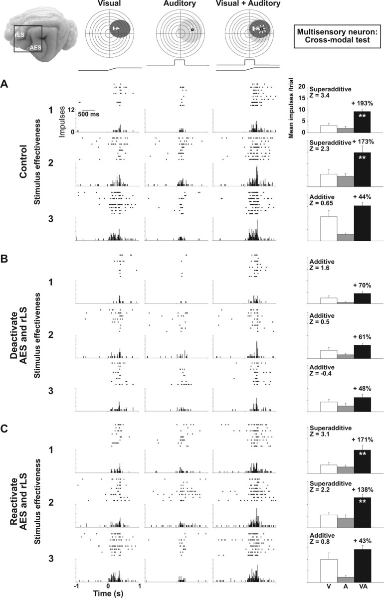
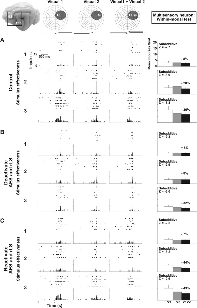
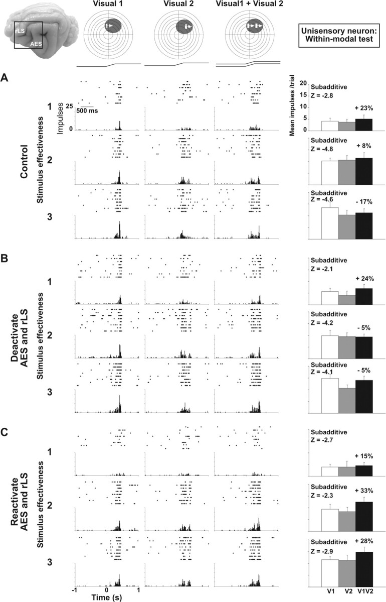
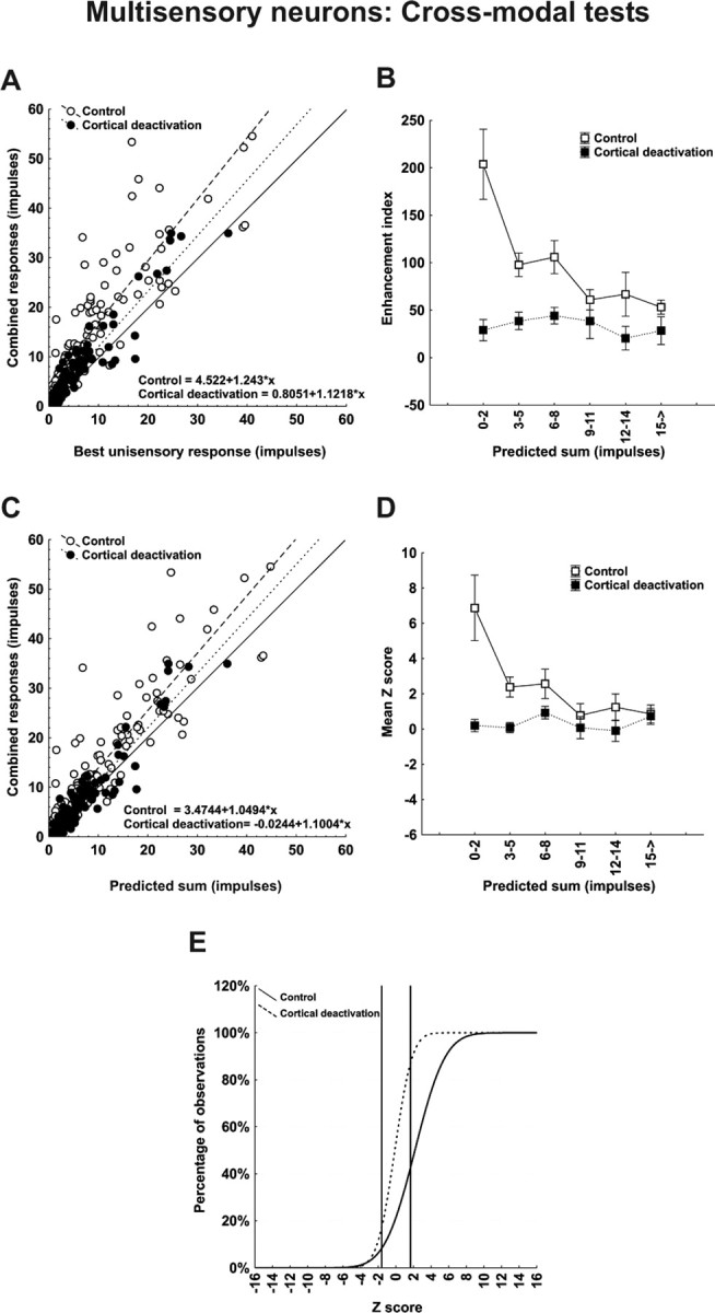
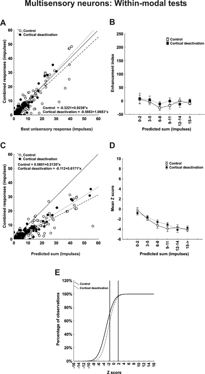
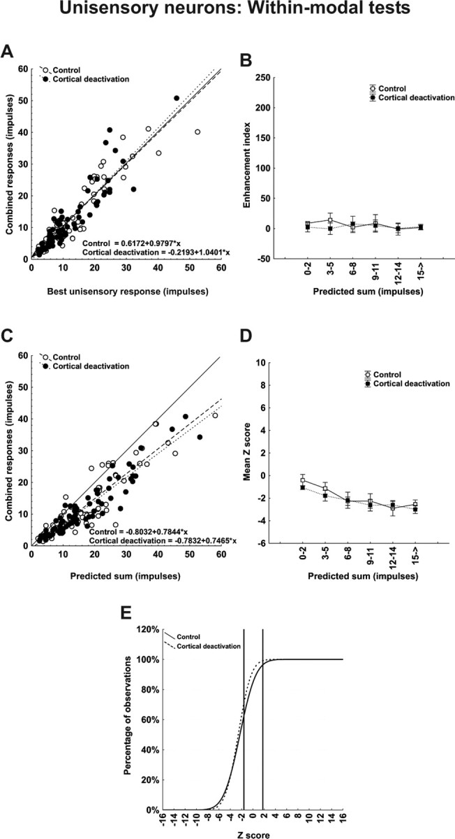
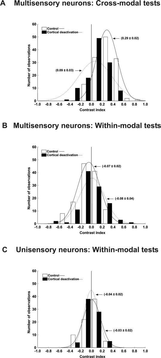
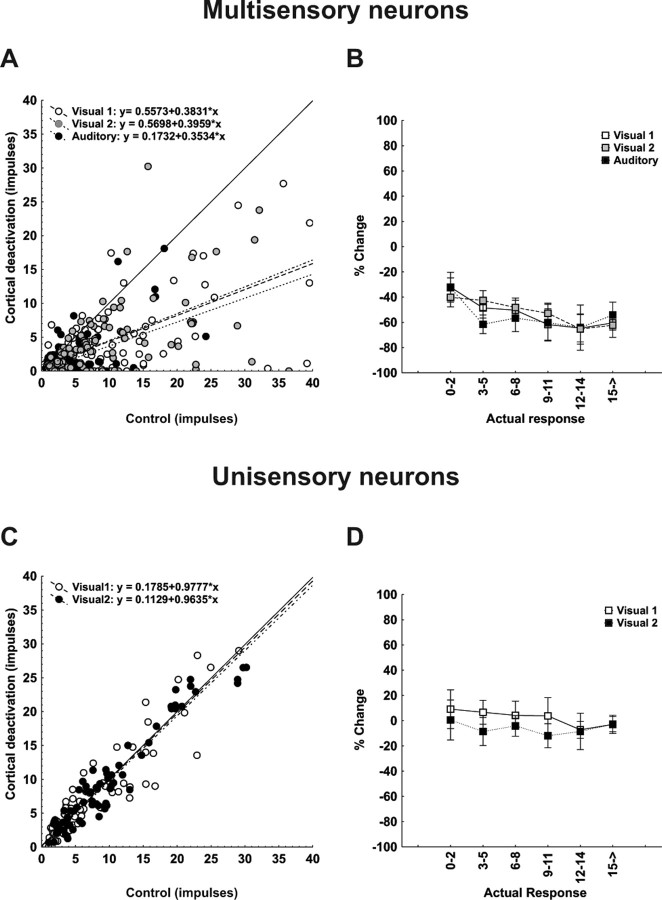
Converging cortical influences from the anterior ectosylvian sulcus and the rostral lateral suprasylvian sulcus were shown to have a multisensory-specific role in the integration of sensory information in superior colliculus (SC) neurons. These observations were based on changes induced by cryogenic deactivation of these cortico-SC projections. Thus, although the results indicated that they played a critical role in integrating SC responses to stimuli derived from different senses (i.e., visual-auditory), they played no role in synthesizing its responses to stimuli derived from within the same sense (visual-visual). This was evident even in the same multisensory neurons. The results suggest that very different neural circuits have evolved to code combinations of cross-modal and within-modal stimuli in the SC, and that the differences in multisensory and unisensory integration are likely caused by differences in the configuration of each neuron's functional inputs rather than to any inherent differences among the neurons themselves. The specificity of these descending influences was also apparent in the very different ways in which they affected responses to the component cross-modal stimuli and their actual integration. Furthermore, they appeared to target only multisensory neurons and not their unisensory neighbors.


Figures

Figure 1.
Figure 1.
Cortical deactivation affects multisensory integration in an SC neuron. At the top are the visual (dark ovoid) and the auditory (light ovoid) receptive fields of this multisensory neuron with icons showing the positions of the visual and auditory stimuli. The moving bar of light (arrow) is indicated by the ramp and the broad-band noise by the square wave above the rasters. The brain photograph shows cortical areas AES and rLS. A–C, Below the receptive fields are shown the neuron's responses to the visual, auditory, and combined visual-auditory stimuli in control (A), cortical deactivation (B), and reactivation conditions (C). Responses are displayed in rasters, histograms and summary bar graphs at three levels of visual stimulus effectiveness. A, Note that multisensory enhancement occurred at every stimulus effectiveness level, but was greatest at the lower end of the neuron's dynamic range. Z scores showed that at this level the enhancement was superadditive, transitioning to additivity at the highest level. B, Cortical deactivation (area in dotted lines) eliminated multisensory enhancement, rendering SC multisensory responses additive at all stimulus effectiveness levels. Modality-specific responses were not significantly affected at lowest level of stimulus effectiveness, but the impact was more evident as the responses increase in intensity. C, Multisensory enhancement was reestablished by cortical reactivation. Impulse counts were taken within a 1 s window. **p < 0.01.
Figure 2.
Figure 2.
Cortical deactivation fails to affect unisensory integration in a multisensory SC neuron. Within-modal visual tests (Visual 1 + Visual 2) were conducted in the same multisensory neuron depicted in Figure 1. A, In the control condition, the neuron showed no response enhancement and an underlying subadditive computation at all levels of stimulus effectiveness. B, Although cortical deactivation reduced the sensory responses (revealing that some of the sensory drive came from cortex), the underlying subadditive computation showed little change. C, Responses were re-established by cortical reactivation. Conventions are the same as in Figure 1.
Figure 3.
Figure 3.
Cortical deactivation fails to affect unisensory integration in a unisensory visual SC neuron. The within-modal visual responses of a unisensory neuron were similar to those of the multisensory neuron in Figure 2. A, In the control condition, the within-modal response was similar to that of the individual component stimuli and was characterized by subadditivity at all levels of stimulus effectiveness. B, C, Cortical deactivation (B) or reactivation (C) had little effect on the modality-specific component responses or the combined within-modal response, and did not alter the underlying subadditive computation. Conventions are the same as in previous figures.
Figure 4.
Figure 4.
Population responses reveal that the computations used by multisensory SC neurons to integrate cross-modal information differ depending on response vigor and that all depend on functional inputs from cortex. A, Each SC neuron's mean response to the cross-modal stimulus combination is plotted against its mean response to the most effective modality-specific component stimulus in both control (open circles) and cortical deactivation (closed circles) conditions. Lines of best fit describe these relationships, and their formulaic descriptions are shown at the bottom of the graph. Note that cortical deactivation shifted the line of best fit toward unity. B, Here, the mean enhancement indices obtained experimentally were parsed into six levels according to the total number of impulses in the multisensory response regardless of the test condition. They were plotted against the predicted mean responses obtained by averaging and summing each neuron's component responses. Note that during the control condition the enhancement index was inversely related to the number of impulses evoked. During cortical deactivation, however, enhancement was rendered minimal and there was no change in the index as a function of impulse number. C, D, These same relationships were noted when plotting combined versus predicted response (C) and mean Z score as a function of predicted sum (D). In the latter case, the superadditive computation obtained when multisensory responses contained low-impulse numbers were lost when responses contained higher impulse numbers, but no superadditivity and no corresponding changes in Z scores were evident during cortical deactivation. E, A cumulative distribution of Z scores shows that ∼60% of the multisensory computations were superadditive in control conditions, but <12% of them were retained during cortical deactivation. They shifted downward to additivity.
Figure 5.
Figure 5.
Population responses reveal that cortex contributes marginally to the vigor with which multisensory SC neurons respond to within-modal stimuli but not at all to crafting the underlying computation. Data were taken from the same neurons used in Figure 4. Here, however, the comparisons are for responses to within-modal stimuli. A, C, Note that the within-modal responses were not significantly different from the most effective (best) unisensory responses (A), and were significantly lower than the predicted sum of the responses to the component stimuli (C). Although the within-modal responses were affected by cortical deactivation, the effect was minimal. B, D, E, Thus, the enhancement index was near or below 0, regardless of the number of impulses obtained for responses in both control and cortical deactivation conditions (B), and Z scores reveal that the underlying computations in both conditions were generally subadditive (D) and their distributions were also very similar (E).
Figure 6.
Figure 6.
Neither the vigor nor the computations used by unisensory SC neurons to integrate within-modal stimuli involve cortex. The within-modal comparisons are identical to those conducted for neurons in Figure 5. A, As in Figure 5, the within-modal responses were not different from their responses to the best unisensory component responses. B, Thus, no enhancement was noted. C–E, The within-modal response was substantially lower than the predicted sum of unisensory component responses (C), and the underlying computation was generally subadditive. However, the absence of any demonstrable effect of cortical deactivation suggests that these neurons do not receive sensory inputs from these cortices.
Figure 7.
Figure 7.
The contrast index reveals inherent differences between multisensory and unisensory neurons. A–C, The graphs illustrate the contrast index for the integration of cross-modal (A) and within-modal (B) information in multisensory neurons and within-modal (C) for the unisensory neurons. A, Although cortical deactivation has a greater affect on the integration of cross-modal information, even during deactivation the majority of cross-modal interactions yielded positive contrast values (filled bars). In contrast, values for within-modal interactions, either for multisensory neurons (B) or unisensory neurons (C), were evenly distributed about zero, indicating that, on average, combined responses did not differ from the best modality-specific responses.
Figure 8.
Figure 8.
Modifications of the individual modality-specific responses during the cortical deactivation. The individual modality-specific responses during the cortical deactivation and in the control condition were plotted together across all levels of stimulus effectiveness. A, During the cross-modal tests, the majority of the modality-specific responses in visual 1, visual 2, and auditory fell below the line of unity during the cortical deactivation. B, There was a slight variation in the percentage of reduction with the stimulus effectiveness. When the actual number of impulses was low, the amount of reduction was smaller, whereas at higher number of impulses the reduction was bigger. Conversely, no apparent modifications in the modality-specific responses were observed in the unisensory neurons. C, D, The responses were clustered around the line of unity and their best fits were indistinguishable from it (C), and the magnitudes of the modifications found in the responses were low (D).

Comment in

References

    1. Alvarado JC, Vaughan JW, Stanford TR, Stein BE. Multisensory versus unisensory integration: contrasting modes in the superior colliculus. J Neurophysiol. 2007;97:3193–3205. - PubMed
    1. Anastasio TJ, Patton PE, Belkacem-Boussaid K. Using Bayes' rule to model multisensory enhancement in the superior colliculus. Neural Comput. 2000;12:1165–1187. - PubMed
    1. Bolognini N, Frassinetti F, Serino A, Ladavas E. “Acoustical vision” of below threshold stimuli: interaction among spatially converging audiovisual inputs. Exp Brain Res. 2004;160:273–282. - PubMed
    1. Calvert GA, Spence C, Stein BE. The handbook of the multisensory processes. Cambridge, MA: MIT; 2004.
    1. Clemo HR, Stein BE. Effects of cooling somatosensory cortex on response properties of tactile cells in the superior colliculus. J Neurophysiol. 1986;55:1352–1368. - PubMed

Publication types

LinkOut - more resources