Comparative Study
. 2007 Nov 21;27(47):13000-11.
doi: 10.1523/JNEUROSCI.3373-07.2007.

Modulation of semaphorin3A activity by p75 neurotrophin receptor influences peripheral axon patterning

Affiliations
Comparative Study

Modulation of semaphorin3A activity by p75 neurotrophin receptor influences peripheral axon patterning

Ayal Ben-Zvi et al. J Neurosci. .

Abstract

The p75 neurotrophin receptor (p75(NTR)) interacts with multiple ligands and coreceptors. It is thought to mediate myelin growth inhibition as part of the Nogo receptor complex, in addition to its other roles. Paradoxically, however, peripheral axons of p75(ExonIII-/-) mutant embryos are severely stunted. This inhibition of axon growth may be a result of neurite elongation defects in p75(NTR) mutant neurons. Here, we show that p75(ExonIII-/-) DRG neurons are hypersensitive to the repellent molecule Semaphorin3A (Sema3A). NGF modulates Sema3A activity equally well in both the p75(NTR) mutant and wild-type neurons, indicating that the hypersensitivity of p75(NTR) mutant neurons is probably not related to their NGF receptor activity. Neuropilin1 and p75(NTR) partially colocalize in DRG growth cones. After Sema3A stimulation, the degree of colocalization is dramatically increased, particularly in clusters associated with Sema3A receptor complex activation. Coimmunoprecipitation studies show that p75(NTR) interacts directly with the Sema3A receptors Neuropilin1 and PlexinA4. When coexpressed with both Neuropilin1 and PlexinA4, p75(NTR) reduces the interaction between these two receptor components. Finally, p75(NTR)/Sema3A double-mutant embryos show growth similar to that observed in Sema3A-null mice. These data indicate that p75(NTR) is an important functional modulator of Sema3A activity and that, in the absence of p75(NTR), oversensitivity to Sema3A leads to severe reduction in sensory innervation. Our results also suggest that while inhibition of p75(NTR) in CNS injury may enhance nerve regeneration resulting from the inhibition of myelin-associated protein, it may also inhibit nerve regeneration through its modulation of Sema3A.


Figures

Figure 1.
Figure 1.
Increased sensitivity of p75NTR−/− DRG neurons to Sema3A repellent activity. A, DRG explants from E12.5 wild-type and p75NTR−/− mutant embryos from the same litters were grown in the presence of 10 ng/ml NGF for 20 h, at the end of which Sema3A was added. Control cultures had no Sema3A added. After an additional incubation period of 40 min with or without Sema3A, the explants were fixed and stained with rhodamine phalloidin. Growth cone collapse results represent the means ± SEM of each treatment with no additional Sem3A (empty bar), 7.5 pm Sema3A (bright gray bar), or 15 pm Sema3A (dark gray bar) in four independent experiments. B, C, DRG explants from E12.5 wild-type, heterozygous, and homozygous p75NTR mutants were grown as in A and treated as described in Materials and Methods. B, Each explant was divided into four quarters as shown in the diagram. An example of one such quarter of an explant at t = 0 and t = 6 is shown. Scale bar, 300 μm. C, Neurite outgrowth assay results represent the means ± SEM of each treatment across three independent experiments (empty bars, no Sema3A; gray bar, 15 pm Sema3A; dark gray bar, 30 pm Sema3A).
Figure 2.
Figure 2.
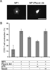
Expression of P75NTR in COS-7 cells reduces Sema3A activity via a PlexinA4/NP1 complex in a concentration-dependent manner. COS-7 cells were transfected with cDNA as indicated and then stimulated with Sema3A (1 nm) 48 h later for 40 min. A, B, Examples of collapsed and noncollapsed transfected cells visualized using GFP expression. Scale bar, 100 μm. C, The percentage of transfected cells that collapsed after Sema3A treatment was determined by fluorescence microscopy. Results represent the means ± SEM of three independent experiments. Cells were transfected with a total of 1 μg cDNA for all treatments. Ten percent of the DNA was pEGFP-N1, 38.5% was PlexinA4, and 26.5% was NP1. p75NTR+ and p75NTR++ indicate that 10 and 26% of the total DNA were p75NTR, respectively. Necl5++ indicates that 26% of the total DNA was a Necl5 expression vector. In each transfection, the total DNA content was brought to 1 μg using pCDNA3 expression vector.
Figure 3.
Figure 3.
Colocalization of NP1 and p75NTR in DRG axons. Sensory axons from E13.5 were stained with anti-p75NTR and anti-NP1 and analyzed by confocal microscopy. Scale bar, 10 μm. For the control experiments, FITC-donkey anti-goat was incubated with rabbit anti-p75-labeled neurons. In addition, Cy3-donkey anti-rabbit was incubated with goat anti-NP1-labeled neurons. Acquisition parameters were defined so that background staining would show no detectable signal (data not shown). Representative optical section images of growth cones with (bottom) or without treatment (top) of 30 pm Sema3A are shown. A clear, but incomplete overlap between p75NTR and NP1 is shown in the image of the overlap pixels (right). After Sema3A treatment, the reorganization of NP1 into clusters is visible (examples of such clusters are indicated by arrows). Quantification of the degree of overlap between NP1/p75 was measured as described in the Materials and Methods section. Each data point represents a mean ± SEM of 30 growth cones from three independent experiments.
Figure 4.
Figure 4.
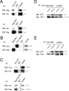
p75NTR immunoprecipitates with NP1 and PlexinA4. A–E, HEK293T cells were transfected with HA-tagged p75NTR and Flag-tagged NP1 (A), myc-Necl5 (nectin-like-5) and Flag-tagged NP1 (B), HA-tagged p75NTR and Flag-PlexinA4 or myc-PlexinA4 (C), myc-PlexinA4 and p75NTR-ECD (extracellular and transmembrane domains) or Flag-p75NTR-ICD (intracellular and transmembrane domains) (D), or myc-NP1 and p75NTR-ECD or Flag-p75NTR-ICD (E). The cells were lysed 48 h after transfection and tested in a coimmunoprecipitation assay. The starting material (lysate) is shown at the right of each panel, the immunoprecipitation antibody is indicated above each lane, and the Western blot (WB) antibody is indicated to the left of each panel. A, Cell lysates were immunoprecipitated with anti-HA-p75 (top) and blotted with anti-Flag, and then with anti-HA-p75NTR. In a separate sample, the cell lysates were immunoprecipitated with anti-Flag-NP1 and blotted with anti-HA, and then with anti-Flag (bottom). Total IgG was also used to immunoprecipitate cell lysates as a control (center lanes). B, Cell lysates were immunoprecipitated with anti-Flag-NP1 or anti-myc-Necl and blotted with anti-Flag or anti-myc as indicated. C, Cell lysates were immunoprecipitated with anti-HA-p75NTR, resulting in immunoprecipitation of myc-PlexinA4 (top). Likewise, immunoprecipitation of Flag-PlexinA4 resulted in immunoprecipitation of HA-p75NTR (bottom). Total IgG failed to immunoprecipitate either HA-p75NTR or Flag-PlexinA4. D, E, Cell lysates were immunoprecipitated with anti-myc-PlexinA4 (D) or anti-myc-NP1 (E) and blotted with anti-p75 extracellular domain antibody in the case of p75NTR-ECD, or anti-Flag in the case of Flag-p75NTR-ICD. To monitor the initial amounts of myc-Plexin A4 (D) or myc-NP1, the membranes were reblotted with anti-myc antibody.
Figure 5.
Figure 5.
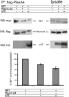
p75NTR neurotrophin receptor disrupts the formation of the Plexin A4/NP1 complex. Top, HEK293T cells were transfected with 4 μg myc-tagged NP1 and 6 μg Flag-tagged PlexinA4. In addition, the cells were transfected with increasing amounts of HA-tagged p75NTR (+ represents 4 μg of p75NTR; ++ represents 10 μg). For all transfections, total DNA was brought to 20 μg using a pCDNA3 expression vector. The cells were lysed at 48 h after transfection and PlexinA4 was immunoprecipitated from lysates with anti-Flag antibodies. Blots were probed with anti-myc-NP1 and then reprobed, first with anti-HA-p75NTR, and then with anti-Flag-PlexinA4. Aliquots of the total cell lysates were directly probed with anti-myc-NP1, anti-Flag-PlexinA4 and anti-HA-p75NTR as a control. (This figure was prepared by combining two parts of the same membrane to exclude a nonrelevant lane). Bottom, Quantification of NP1 coimmunoprecipitated by PlexinA4. We normalized the levels of NP1 and PlexinA4 to their expression levels in the lysate. We then divided the normalized values of NP1 by the normalized values of PlexinA4. The result of this calculation in the absence of p75 was defined as 100% coimmunoprecipitation. Results represent the means ± SEM of five independent experiments.
Figure 6.
Figure 6.
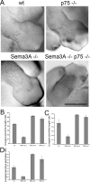
Growth inhibition of peripheral sensory projections in p75NTR mutant embryos is alleviated in the absence of Sema3A. Whole-mount anti-neurofilament immunohistochemistry was performed on E12.5 embryos. A, Peripheral nerves in hind limbs. Peripheral nerves in p75NTR mutant mice are severely stunted (top right), compared with those of control wild-type littermates (top left). In contrast, in p75NTR mutant mice that were also Sema3A-null (bottom right), peripheral axons overshot the front observed in wild-type embryos, similar to the overshooting observed in Sema3A-null mice that are wild-type for the p75NTR gene (bottom left). Scale bar, 1 mm. B–D, Quantification of relative innervation of hind limbs (representative whole mount shown in A) forelimbs (whole mount shown in S3) and trigeminal ganglia (whole mount shown in S4) using whole-mount anti-neurofilament immunohistochemistry. To quantify the axon growth capacity of each genotype, we measured the length of the longest axon. B, C, The length of the longest axon measured from the base of the limb and normalized to the length of the hind limb (B) or forelimb (C). D, The length of the longest axon from the trigeminal ganglion normalized to the eye perimeter. Results represent the means ± SEM of seven wild-type embryos, 15 Sema3A mutants, four p75NTR mutants and five Sema3A/p75NTR double-mutant embryos from a total of eight litters.
Figure 7.
Figure 7.
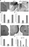
Growth inhibition of peripheral sensory projections in p75NTR mutant mice is sensitive to changes in Sema3A levels. Whole-mount anti-neurofilament immunohistochemistry was performed on E12.5 embryos. A, D, Peripheral nerves in forelimbs (A) and trigeminal ganglia (D) are shown. Compared with those of the control wild-type littermates (left), peripheral nerves in p75NTR mutant mice (middle) were severely stunted. In contrast, p75NTR−/−, Sema3A+/− mutant mice (right) resemble their wild-type littermates. B, C, E, Quantification of relative innervation of forelimbs (A), trigeminal ganglia (D) and hind limbs (S5) using whole-mount anti-neurofilament immunohistochemistry. We again used the longest axon length normalized to the length of the limb (B, C) or the eye perimeter (E) to quantify the axon growth capacity of each genotype. Forelimbs (B), hind limbs (C), and trigeminal ganglion (E) are shown. F, Quantification of total axon lengths in forelimbs (gray bars) and hind limbs (empty bars) was estimated using Buffon's needle problem equation (see Materials and Methods). Results represent the means ± SEM of seven wild-type, three p75NTR+/−, four p75NTR−/− mutant (wild type and p75NTR−/− mutant are the same embryos used for Fig. 6), and seven Sema3A+/−, p75NTR−/− embryos. Scale bar, 1 mm.

References

    1. Bandtlow C, Dechant G. From cell death to neuronal regeneration, effects of the p75 neurotrophin receptor depend on interactions with partner subunits. Sci STKE. 2004;2004:pe24. - PubMed
    1. Behar O, Golden JA, Mashimo H, Schoen FJ, Fishman MC. Semaphorin III is needed for normal patterning and growth of nerves, bones and heart. Nature. 1996;383:525–528. - PubMed
    1. Behar O, Mizuno K, Badminton M, Woolf CJ. Semaphorin 3A growth cone collapse requires a sequence homologous to tarantula hanatoxin. Proc Natl Acad Sci USA. 1999;96:13501–13505. - PMC - PubMed
    1. Bentley CA, Lee KF. p75 is important for axon growth and Schwann cell migration during development. J Neurosci. 2000;20:7706–7715. - PMC - PubMed
    1. Ben-Zvi A, Yagil Z, Hagalili Y, Klein H, Lerman O, Behar O. Semaphorin 3A and neurotrophins: a balance between apoptosis and survival signaling in embryonic DRG neurons. J Neurochem. 2006;96:585–597. - PubMed

Publication types

MeSH terms

LinkOut - more resources