Comparative Study
. 2007 Nov 21;27(47):13033-41.
doi: 10.1523/JNEUROSCI.4290-06.2007.

Toll-like receptor 3 is a potent negative regulator of axonal growth in mammals

Affiliations
Comparative Study

Toll-like receptor 3 is a potent negative regulator of axonal growth in mammals

Jill S Cameron et al. J Neurosci. .

Abstract

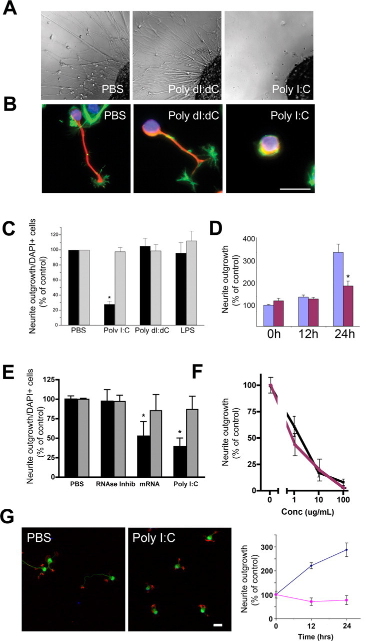
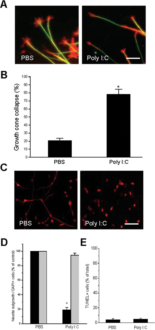
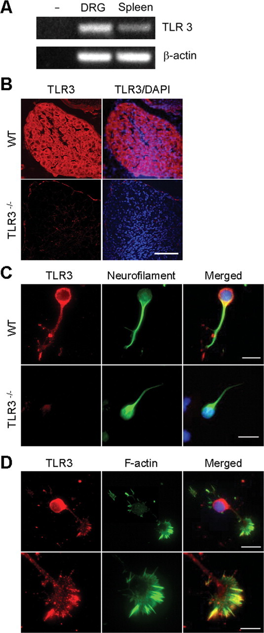
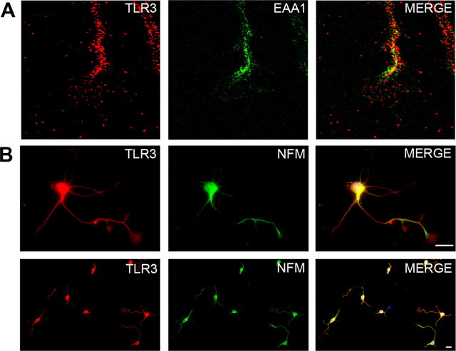
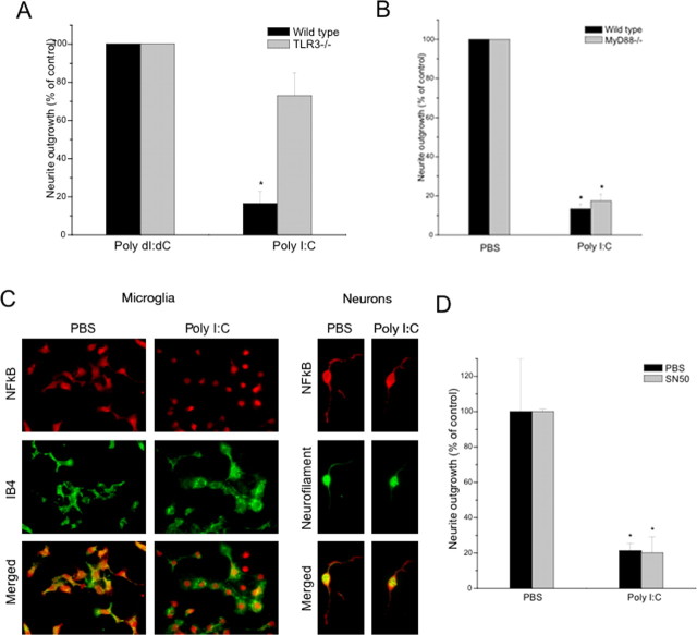
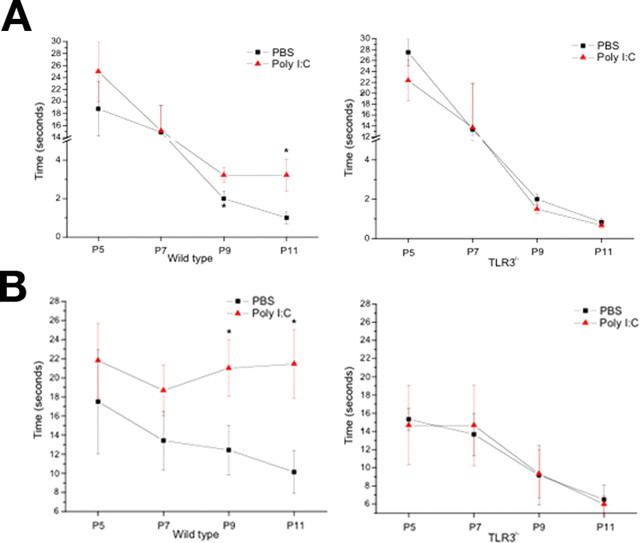
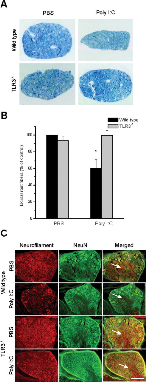
Toll is a cell surface receptor with well described roles in the developmental patterning of invertebrates and innate immunity in adult Drosophila. Mammalian toll-like receptors represent a family of Toll orthologs that function in innate immunity by recognizing molecular motifs unique to pathogens or injured tissue. One member in this family of pattern recognition receptors, toll-like receptor 3 (TLR3), recognizes viral double-stranded RNA and host mRNA. We examined the expression and function of TLRs in the nervous system and found that TLR3 is expressed in the mouse central and peripheral nervous systems and is concentrated in the growth cones of neurons. Activation of TLR3 by the synthetic ligand polyinosine:polycytidylic acid (poly I:C) or by mRNA rapidly causes growth cone collapse and irreversibly inhibits neurite extension independent of nuclear factor kappaB. Mice lacking functional TLR3 were resistant to the neurodegenerative effects of poly I:C. Neonatal mice injected with poly I:C were found to have fewer axons exiting dorsal root ganglia and displayed related sensorimotor deficits. No effect of poly I:C was observed in mice lacking functional TLR3. Together, these findings provide evidence that an innate immune pattern recognition receptor functions autonomously in neurons to regulate axonal growth and advances a novel hypothesis that this class of receptors may contribute to injury and limited CNS regeneration.


Figures

Figure 1.
Figure 1.
Poly I:C is a negative regulator of neurite outgrowth. A, Differential interference contrast images of E9 chick DRG explants cultured overnight in the presence of the TLR3 agonist poly I:C (20 μg/ml) or relevant controls, poly dI:dC (20 μg/ml), or PBS. B, Neurofilament (red) and f-actin (green) staining of E14 mouse sensory neurons cultured for 4 h in the presence of poly I:C, poly dI:dC, or PBS. Scale bar, 25 μm. C, Quantification of neurite outgrowth (black bars) and DAPI staining (gray bars) from B in percent for both measures. D, Quantification (percent) of neurite outgrowth of mouse DRGs exposed to poly I:C (20 μg/ml) (red bars) or vehicle control (purple bars) for 0, 12, or 24 h. Cells were grown for 1 d before exposure. E, Quantification of neurite outgrowth (black bars) and DAPI staining (gray bars) from mouse sensory neurons cultured for 4 h in the presence of poly I:C, poly(A)+ mRNA, RNase inhibitor, or PBS. F, Dose–response curve for poly I:C (black line) and RNA (purple line) effects on neurite outgrowth of mouse sensory neurons. G, Neurofilament (green) and f-actin (red) staining of E18 mouse hippocampal neurons cultured for 24 h in the presence of poly I:C (20 μg/ml) or PBS control. Scale bar, 25 μm. Graph at right represents time course of neurite length of hippocampal neurons exposed to poly I:C (purple line) or PBS (blue line). Error bars in this and subsequent figures represent SEM. *p < 0.01, Student's t test.
Figure 2.
Figure 2.
Poly I:C causes growth cone collapse of existing neurites and inhibition of neurite outgrowth is not reversible within 24 h. Explants of E9 chick DRGs were plated overnight and exposed to either PBS or poly I:C (20 μg/ml) for 30 min. A, Immunofluorescent staining of f-actin (red) and neurofilament (green) in DRG explants shows growth-cone collapse as a result of exposure to poly I:C. Scale bar, 25 μm. B, Quantification of explants assayed for growth-cone collapse. Exposure to poly I:C for 30 min caused a significant increase in the total number of collapsed growth cones in DRG explants. C, Neurofilament staining of a representative field of chick sensory neurons treated acutely with PBS or poly I:C (20 μg/ml) for 4 h, washed, then grown overnight in DMEM plus NGF. Scale bar, 100 μm. D, Quantification of the effects of poly I:C on neurite outgrowth (black bars) and cell survival (gray bars) 24 h after 4 h treatment. E, Quantification of TUNEL+ neurons from D. *p < 0.01, Student's t test.
Figure 3.
Figure 3.
TLR3 transcripts and protein are expressed in mouse DRG neurons and colocalize within the growth cone. A, Total RNA was extracted from E14 mouse DRG or adult spleen. TLR3 and actin transcripts were identified by RT-PCR. TLR3 transcripts were detected in DRG and spleen extracts, but not in the absence of RNA. B, Confocal images of immunofluorescent staining for TLR3 (red) and DAPI (blue) in wild-type and TLR3−/− mouse DRGs in situ. Scale bar, 40 μm. C, TLR3 (red) and neurofilament (green) staining of dissociated DRG neurons isolated from E14 wild-type and TLR3−/− embryos. Scale bars: 25 μm. D, TLR3 (red) and f-actin (green) staining of dissociated DRG neurons and growth cones. Scale bars: top, 25 μm; bottom, 10 μm.
Figure 4.
Figure 4.
TLR3 is partly endosomal and found in multiple neuronal subtypes. A, Confocal imaging of a mouse DRG growth cone containing TLR3 (red) and the endosomal marker, EAA1 (green). B, E17 cortical neurons at high magnification (top) and low magnification (bottom) stained with TLR3 (red) and neurofilament-M (NFM) (green). Scale bar, 25 μm.
Figure 5.
Figure 5.
Poly I:C inhibits neurite outgrowth through a TLR3-dependent NFκB- and MyD88-independent pathway. A, Quantification of neurite outgrowth from wild-type and TLR3−/− mouse sensory neurons cultured for 4 h in the presence of poly I:C (20 μg/ml) or poly dI:dC (20 μg/ml), as a negative control. No differences in plating density or cell survival were observed by DAPI staining. B, MyD88 loss-of-function has no impact on poly I:C-mediated inhibition of neurite outgrowth. Quantification of neurite outgrowth from wild-type and MyD88−/− sensory neurons cultured for 4 h in the presence of poly I:C (20 μg/ml) or PBS, as a negative control is shown. C, Nuclear translocation of the p65 subunit of NF-κB occurs in microglia but not neurons after treatment with poly I:C. Immunofluorescent staining of NF-κB localization in microglia and neurons after 1 h exposure to poly I:C (20 μg/ml) or PBS. D, Quantification of neurite outgrowth from sensory neurons cultured for 4 h in the presence of poly I:C (20 μg/ml) or PBS with or without the addition of SN50 (50 μg/ml), an inhibitor of NFκB nuclear translocation. *p < 0.01, Student's t test.
Figure 6.
Figure 6.
A, B, Effects of poly I:C (3 mg/kg) or PBS intrathecal injection on righting reflex (A) and negative geotaxis (B) in wild-type and TLR3−/− neonatal mice. P4 neonatal mice were injected intrathecally with 3 μl of either PBS or poly I:C (2 mg/ml) at a final concentration of 3 μg/ml. Behavioral testing was performed at P5, P7, P9, and P11. Righting reflex and negative geotaxis tasks were conducted and scored as described in Materials and Methods. *p < 0.01.
Figure 7.
Figure 7.
Intrathecal injection of poly I:C inhibits the normal development of sensory neurons in vivo via TLR3. A, Cross-sections of dorsal roots isolated from wild-type and TLR3−/− mice injected intrathecally with poly I:C (3 mg/kg) or PBS. Representative images are from the L3 dorsal root of each animal. B, Quantification of the number of myelinated fibers in dorsal root cross sections for L1–L5. C, Neurofilament (red) and NeuN (green) immunofluorescent images of DRGs from animals in A. Note the smaller ganglion size and lack of neurofilament intensity in ganglion roots (arrows) in wild-type animals injected with poly I:C, whereas there is no change in size or neurofilament staining in the other groups, as expected. Scale bar, 50 μm.

References

    1. Alexopoulou L, Holt AC, Medzhitov R, Flavell RA. Recognition of double-stranded RNA and activation of NF-κB by Toll-like receptor 3. Nature. 2001;413:732–738. - PubMed
    1. Andaloussi AE, Sonabend AM, Han Y, Lesniak MS. Stimulation of TLR9 with CpG ODN enhances apoptosis of glioma and prolongs the survival of mice with experimental brain tumors. Glia. 2006;54:526–535. - PubMed
    1. Anderson KV, Jurgens G, Nusslein-Volhard C. Establishment of dorsal-ventral polarity in the Drosophila embryo: genetic studies on the role of the Toll gene product. Cell. 1985a;42:779–789. - PubMed
    1. Anderson KV, Bokla L, Nusslein-Volhard C. Establishment of dorsal-ventral polarity in the Drosophila embryo: the induction of polarity by the Toll gene product. Cell. 1985b;42:791–798. - PubMed
    1. Bergmann-Leitner ES, Leitner WW. Danger, death and DNA vaccines. Microbes Infect. 2004;6:319–327. - PubMed

Publication types

MeSH terms

LinkOut - more resources