. 2008 Jan;18(1):161-71.
doi: 10.1101/gr.7020108. Epub 2007 Nov 21.

Chlamydia trachomatis: genome sequence analysis of lymphogranuloma venereum isolates

Affiliations

Chlamydia trachomatis: genome sequence analysis of lymphogranuloma venereum isolates

Nicholas R Thomson et al. Genome Res. 2008 Jan.

Abstract

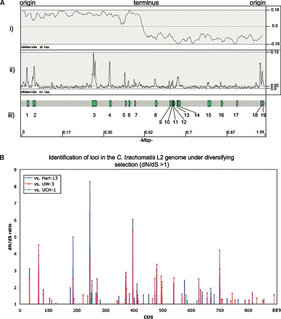
Chlamydia trachomatis is the most common cause of sexually transmitted infections in the UK, a statistic that is also reflected globally. There are three biovariants of C. trachomatis: trachoma (serotypes A-C) and two sexually transmitted pathovars; serotypes D-K and lymphogranuloma venereum (LGV). Trachoma isolates and the sexually transmitted serotypes D-K are noninvasive, whereas the LGV strains are invasive, causing a disseminating infection of the local draining lymph nodes. Genome sequences are available for single isolates from the trachoma (serotype A) and sexually transmitted (serotype D) biotypes. We sequenced two isolates from the remaining biotype, LGV, a long-term laboratory passaged strain and the recent "epidemic" LGV isolate-causing proctitis. Although the genome of the LGV strain shows no additional genes that could account for the differences in disease outcome, we found evidence of functional gene loss and identified regions of heightened sequence variation that have previously been shown to be important sites for interstrain recombination. We have used new sequencing technologies to show that the recent clinical LGV isolate causing proctitis is unlikely to be a newly emerged strain but is most probably an old strain with relatively new clinical manifestations.


Figures

Figure 1.
Figure 1.
Circular representation of the C. trachomatis strain L2 chromosome. The outer scale shows the size in base pairs. From the outside in, circles 1 and 2 show the position of CDSs transcribed in a clockwise and anticlockwise direction. Using the published gene predictions for C. trachomatis strains UW-3 and Har-13, the strain L2 CDSs have been color-coded depending on whether they are: (blue) predicted and intact in all isolates; (pink) predicted and intact in L2 and UW-3; (green) predicted and intact in L2 and Har-13; (orange) defunct in L2, predicted and intact in Har-13 and UW-3; (red) unique to L2; (brown) defunct in all isolates. (Circles 3–10) C. trachomatis strain L2 CDSs that are present/absent in: C. muridarum (circles 3 and 4; present gray; absent yellow), Cp. felis (circles 5 and 6; present green; absent pink), Cp. caviae (circles 7 and 8; present pink; absent blue), and Cp. pneumoniae (circles 9 and 10; present red; absent blue) by reciprocal FASTA analysis. Circle 11 shows a plot of G+C content (in a 0.5-kb window); circle 12 shows a plot of GC skew ([G-C]/[G+C]; in a 0.5-kb window). The position of the PZ (pink) and the PLD CDSs in locus 1 and 3 are numbered accordingly and marked (red). Since the gene content of strain L2 and UCH-1 are essentially identical, these data apply equally to both isolates.
Figure 2.
Figure 2.
Distribution of predicted and functional CDSs shared between C. trachomatis strains L2, UW-3, and Har-13. The Venn diagram shows the number of CDS predicted to be functional, that are unique or shared, between one or more C. trachomatis isolates (see Methods). Pseudogenes were counted as absent in this analysis.
Figure 3.
Figure 3.
Comparison of the PZ locus of C. trachomatis strains L2, UW-3, and Har-13 ACT comparison (http://www.sanger.ac.uk/Software/ACT) of amino acid matches between the complete six-frame translations (computed using TBLASTX) of representatives of the PZ regions of the three sequenced C. trachomatis genomes: Har-13, C. trachomatis strain Har-13; UW-3, C. trachomatis strain UW-3; and L2, C. trachomatis strain L2. The red bars spanning between the genomes represent individual TBLASTX matches. Forward and reverse strands of DNA are shown for each genome (dark-gray lines). CDS are marked as colored boxes positioned on the three forward and three reverse translation reading frames (pale-gray lines). Regions mentioned in the text are marked and the CDSs labeled and color coded: Cytotoxin gene fragments (yellow), tryptophan biosynthetic genes (intact pale green; pseudogene orange), and phospholipase D (Locus 2; orthologous PLD CDSs are colored similarly). PZ regions are marked (purple box on DNA lines). The position of deletion events (*) and frameshift mutations (black arrowheads) within the PLD CDS are marked. Multiple systematic gene identifiers contained within parentheses indicates that the CDS was originally annotated as two CDSs.
Figure 4.
Figure 4.
Phylogenetic relationships of chlamydial phospholipase D proteins. The protein names have been colored to indicate chlamydial strains: C. trachomatis strains: L2 (pink), UW-3 (red), and Har-13 (green); C. muridarum (dark blue); Cp. felis (light blue); Cp. caviae (yellow); Cp. abortus (orange); Cp. pneumoniae (brown); Candidatus Protochlamydia amoebophila UWE25 (black). Maximum likelihood tree built from protein sequences, using ClustalX, Phylip (Version 3.6), and NJplot. The numbers at the tree branches are percentage bootstrap values indicating the confident levels at that node where congruent. The bar indicates the genetic distance between species (one substitution per 100) as displayed in the branch lengths.
Figure 5.
Figure 5.
(A) Distribution of SNPs over the C. trachomatis strain L2 genome compared with strains Har-13, UW-3, and UCH-1. (i) The G-C skew (G+C/G-C). The position of the origin and terminus are marked. (ii) Shows the sum of all the SNPs in C. trachomatis strain L2 compared with strains Har-13, UW-3, and UCH-1, plotted as a frequency over a given window size. (iii) Regions with a high SNP density are numbered and the CDSs within this region are described in Supplemental Table 3. The base-pair positions are given in Mbp (bottom) and the window sizes are marked. The maximum, minimum, and average frequencies for each plot are given, where appropriate (right). (B) The distribution of C. trachomatis strain L2 CDSs with a dN/dS ratio >1. The dN/dS values for all of the C. trachomatis strain L2 CDSs (x axis) compared with their orthologs in strains Har-13, UW-3, and UCH-1 are shown (dN/dS >1 only). CDS displaying a high dN/dS ratio are described in Table 3.

References

    1. Azuma Y., Hirakawa H., Yamashita A., Cai Y., Rahman M.A., Suzuki H., Mitaku S., Toh H., Goto S., Murakami T., Hirakawa H., Yamashita A., Cai Y., Rahman M.A., Suzuki H., Mitaku S., Toh H., Goto S., Murakami T., Yamashita A., Cai Y., Rahman M.A., Suzuki H., Mitaku S., Toh H., Goto S., Murakami T., Cai Y., Rahman M.A., Suzuki H., Mitaku S., Toh H., Goto S., Murakami T., Rahman M.A., Suzuki H., Mitaku S., Toh H., Goto S., Murakami T., Suzuki H., Mitaku S., Toh H., Goto S., Murakami T., Mitaku S., Toh H., Goto S., Murakami T., Toh H., Goto S., Murakami T., Goto S., Murakami T., Murakami T., et al. Genome sequence of the cat pathogen, Chlamydophila felis. DNA Res. 2006;13:15–23. - PubMed
    1. Bannantine J.P., Rockey D.D., Hackstadt T., Rockey D.D., Hackstadt T., Hackstadt T. Tandem genes of Chlamydia psittaci that encode proteins localized to the inclusion membrane. Mol. Microbiol. 1998a;28:1017–1026. - PubMed
    1. Bannantine J.P., Stamm W.E., Suchland R.J., Rockey D.D., Stamm W.E., Suchland R.J., Rockey D.D., Suchland R.J., Rockey D.D., Rockey D.D. Chlamydia trachomatis IncA is localized to the inclusion membrane and is recognized by antisera from infected humans and primates. Infect. Immun. 1998b;66:6017–6021. - PMC - PubMed
    1. Bannantine J.P., Griffiths R.S., Viratyosin W., Brown W.J., Rockey D.D., Griffiths R.S., Viratyosin W., Brown W.J., Rockey D.D., Viratyosin W., Brown W.J., Rockey D.D., Brown W.J., Rockey D.D., Rockey D.D. A secondary structure motif predictive of protein localization to the chlamydial inclusion membrane. Cell. Microbiol. 2000;2:35–47. - PubMed
    1. Behets F.M., Andriamiadana J., Randrianasolo D., Randriamanga R., Rasamilalao D., Chen C.Y., Weiss J.B., Morse S.A., Dallabetta G., Cohen M.S., Andriamiadana J., Randrianasolo D., Randriamanga R., Rasamilalao D., Chen C.Y., Weiss J.B., Morse S.A., Dallabetta G., Cohen M.S., Randrianasolo D., Randriamanga R., Rasamilalao D., Chen C.Y., Weiss J.B., Morse S.A., Dallabetta G., Cohen M.S., Randriamanga R., Rasamilalao D., Chen C.Y., Weiss J.B., Morse S.A., Dallabetta G., Cohen M.S., Rasamilalao D., Chen C.Y., Weiss J.B., Morse S.A., Dallabetta G., Cohen M.S., Chen C.Y., Weiss J.B., Morse S.A., Dallabetta G., Cohen M.S., Weiss J.B., Morse S.A., Dallabetta G., Cohen M.S., Morse S.A., Dallabetta G., Cohen M.S., Dallabetta G., Cohen M.S., Cohen M.S. Chancroid, primary syphilis, genital herpes, and lymphogranuloma venereum in Antananarivo, Madagascar. J. Infect. Dis. 1999;180:1382–1385. - PubMed

Publication types

MeSH terms

LinkOut - more resources