. 2007 Dec 12;26(24):5153-66.
doi: 10.1038/sj.emboj.7601918. Epub 2007 Nov 22.

The structural basis of cyclic diguanylate signal transduction by PilZ domains

Affiliations

The structural basis of cyclic diguanylate signal transduction by PilZ domains

Jordi Benach et al. EMBO J. .

Abstract

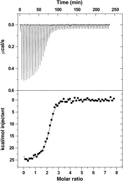
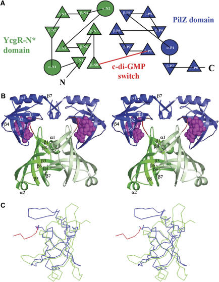
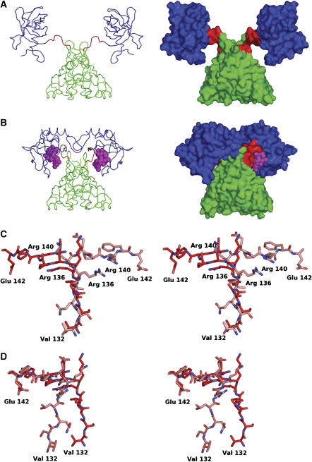
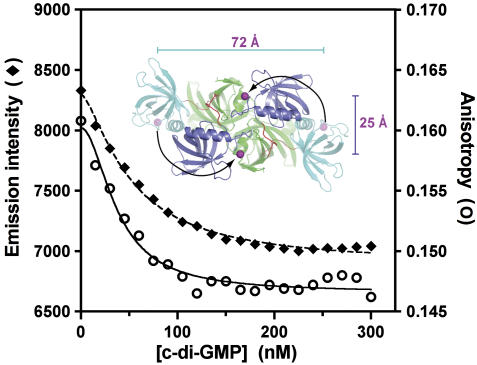
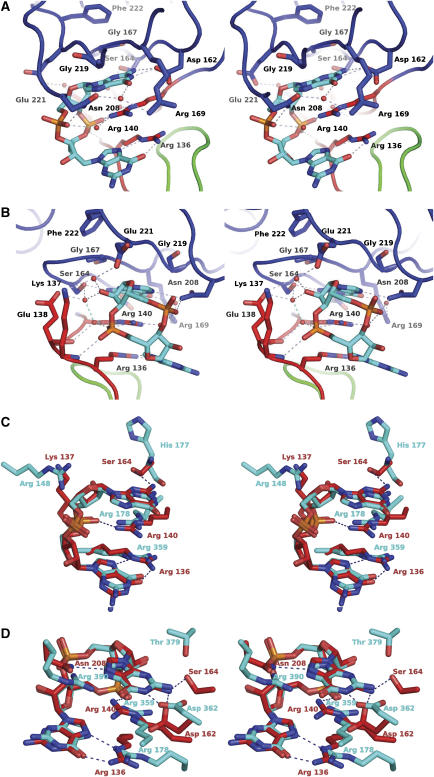
The second messenger cyclic diguanylate (c-di-GMP) controls the transition between motile and sessile growth in eubacteria, but little is known about the proteins that sense its concentration. Bioinformatics analyses suggested that PilZ domains bind c-di-GMP and allosterically modulate effector pathways. We have determined a 1.9 A crystal structure of c-di-GMP bound to VCA0042/PlzD, a PilZ domain-containing protein from Vibrio cholerae. Either this protein or another specific PilZ domain-containing protein is required for V. cholerae to efficiently infect mice. VCA0042/PlzD comprises a C-terminal PilZ domain plus an N-terminal domain with a similar beta-barrel fold. C-di-GMP contacts seven of the nine strongly conserved residues in the PilZ domain, including three in a seven-residue long N-terminal loop that undergoes a conformational switch as it wraps around c-di-GMP. This switch brings the PilZ domain into close apposition with the N-terminal domain, forming a new allosteric interaction surface that spans these domains and the c-di-GMP at their interface. The very small size of the N-terminal conformational switch is likely to explain the facile evolutionary diversification of the PilZ domain.


Figures

Figure 1a
Figure 1a
Phylogenetic analysis of eubacterial PilZ domains. These analyses were modeled on those of Amikam and Galperin (2006). (A) Cladogram showing evolutionary relationships between PilZ domains inferred from maximum parsimony analysis of a multiple sequence alignment (containing only the PilZ domain from each protein). The name of a representative member of each functional protein family is given, with the first two letters representing the SwissProt species code and the open-reading-frame number used for proteins lacking functional annotation. The number of likely orthologs identified in 295 fully sequenced eubacterial genomes is indicated in parentheses, while the diagrams schematize the overall domain organization inferred using RPS-BLAST analysis of the Conserved Domain Database (CDD) (Marchler-Bauer et al, 2005). Numbers at internal nodes in the tree represent the support for a split at the indicated evolutionary distance (but are not shown for 100% support). Protein domains are labeled as follows: CelS, cellulose synthetase domain; EAL, c-di-GMP phosphodiesterase domain; GGDEF, c-di-GMP cyclase domain; GlyT2, glycosyl transferase domain; MdoH, domain of membrane glycosyltransferases in COG2943; YcgR-N, strong sequence homology to the N-terminal domain of the E. coli YcgR protein at a level equivalent to that found in proteins in COG5581; YcgR-N*, weaker sequence homology to the YcgR-N domain; ???, uncharacterized conserved domain. The single PilZ-domain proteins DgrA and DgrB from C. crescentus are not represented here because likely orthologous proteins were not identified in other organisms.
Figure 1b
Figure 1b
(B) Sequence–structure alignment generated by ESPRIPT (Gouet et al, 1999). Arrows represent β-strands and coils represent α-helices. Secondary structural elements are numbered independently in the two domains and colored according to domain of origin, with the YcgR-N* domain green, the c-di-GMP switch red, and the remainder of the PilZ domain blue. Magenta symbols represent van der Waals contacts to c-di-GMP, with closed circles and open squares indicating the presence or absence of H-bonds, respectively. The sequence alignment, which has the most strongly conserved sites highlighted in red, includes the other PilZ-domain-containing proteins of known structure plus one representative from each functional protein family in (B) whose domain organization is equivalent to that of VCA0042/PlzD. These were aligned initially using a position-specific score matrix (Altschul et al, 1997) derived from the alignment of Amikam and Galperin (2006) but manually adjusted to reflect the structural alignment of the individual domains.
Figure 2
Figure 2
Isothermal titration calorimetry shows that c-di-GMP binds to VCA0042/PlzD with sub-micromolar affinity. The top trace shows baseline-corrected data collected at 25°C in binding buffer (5 mM MgCl2, 10 mM KCl, 300 mM NaCl, 10% glycerol, 8 mM β-mercaptoethanol, and 10 mM Tris-Cl, pH 8.0), while the bottom trace shows the integrated heat released during each injection as a function of the molar ratio of c-di-GMP to VCA0042/PlzD dimer. The dotted line shows the results of curve fitting using a two-site sequential binding model with c-di-GMP concentration adjusted to give a binding stoichiometry of ∼1. (See page 9 in the Supplementary data for a detailed explanation.) Supplementary Table S1 gives the thermodynamic parameters estimated from these data using a variety of curve-fitting procedures, showing that c-di-GMP binds with affinity better than 350 nM (probably ∼100 nM) in an enthalpically favorable (ΔH<−12 kcal/mol) but entropically unfavorable (TΔS<−3 kcal/mol) reaction.
Figure 3
Figure 3
Crystal structure of the VCA0042/PlzD complex with c-di-GMP. In all panels in this paper, the YcgR-N* domain is colored green, the c-di-GMP switch red, and the remainder of the PilZ domain blue. (A) Topology diagram generated by TOPS (Michalopoulos et al, 2004), with circles and triangles respectively representing α-helices and β-strands running roughly perpendicular to the plane of the page. Connecting segments penetrating into the symbols pass above the plane while those stopping at the boundary of the symbol pass below. The β-strands make exclusively antiparallel H-bonding interactions. (B) Stereo ribbon diagram of the protein dimer with c-di-GMP shown in magenta space-filling representation. Darker or lighter colors are used to distinguish the two subunits in the dimer. Secondary structural elements are numbered separately in the two domains like in Figure 1B. (C) Cα traces showing structural superposition of the YcgR-N* domain and the PilZ domain performed using DALI (Holm and Sander, 1993).
Figure 4
Figure 4
Comparison of apo and c-di-GMP complex structures of VCA0042/PlzD. (A) Cα trace (left) and molecular surface (right) of the apo structure (PBD ID 1YLN; R Zhang, M Zhou, S Moy, F Collart, and A Joachimiak) colored as in Figure 3. (B) Equivalent representations of the c-di-GMP complex structure. (C, D) The alternative conformations of the c-di-GMP switch (residues 134–140) are shown in ball-and-stick representation following least-squares alignment of the Cα atoms in either the N-terminal YcgR-N* domain (C) or the C-terminal PilZ domain (D). Carbon atoms in the apo and complex structures are colored lighter and darker shade of red, respectively. Nitrogen atoms are colored blue and oxygen atoms are colored red.
Figure 5
Figure 5
Fluorescence resonance energy transfer experiments support a change in relative orientation of the C-terminal PilZ domains in VCA0042/PlzD upon c-di-GMP binding. Total fluorescence and anisotropy (measured as described on page 11 in the Supplementary data) are shown for an Alexa-Fluor 488 dye covalently bound to residue 247 in VCA0042/PlzD (in a C207A/N247C double mutant protein). The magenta spheres in the inset show the separations of the Cα atoms of residue 247 in the protein dimers observed in the apo (72 Å) and c-di-GMP-bound (25 Å) crystal structures of the wild-type protein. The experiment was conducted in binding buffer at 25°C (like the ITC experiment in Figure 2).
Figure 6
Figure 6
Stereochemistry of the c-di-GMP-binding site in VCA0042/PlzD.The stereo pairs have nitrogen, oxygen, and phosphorus atoms colored blue, red, and orange, respectively. Dotted lines indicate H-bonds. (A, B) Two views of the c-di-GMP-binding site in the complex structure. Carbon atoms and backbone worms are colored according to domain/region of origin (green for the YcgR-N* domain, red for the c-di-GMP switch, and blue for the C-terminal PilZ domain). A subset of the ordered water molecules in the cooperative H-bonding network is shown. See also Supplementary Figure S6. (C, D) Least-squares superposition of the c-di-GMP molecule in VCA0042/PlzD with each of the two different c-di-GMP molecules bound in the allosteric regulatory site of the PleD guanylate cyclase (PDB ID 1W25) (Chan et al, 2004). Carbon atoms and residue numbers from PleD or VCA0042 are colored cyan or red, respectively. The c-di-GMP molecules from PleD shown in these two panels interact with each other to form a base-stacked dimer (illustrated in context in the PleD structure in Supplementary Figure S7). Therefore, many of the PleD side chains shown in these two panels are the same, including the two arginine residues forming the dual guanidino motif.

References

    1. Ades SE (1995) The engrailed homeodomain: determinants of DNA-binding affinity and specificity. PhD Thesis. Department of Biology, Massachusetts Institute of Technology, Cambridge, MA
    1. Alm RA, Bodero AJ, Free PD, Mattick JS (1996) Identification of a novel gene, pilZ, essential for type 4 fimbrial biogenesis in Pseudomonas aeruginosa. J Bacteriol 178: 46–53 - PMC - PubMed
    1. Altschul SF, Madden TL, Schaffer AA, Zhang J, Zhang Z, Miller W, Lipman DJ (1997) Gapped BLAST and PSI-BLAST: a new generation of protein database search programs. Nucleic Acids Res 25: 3389–3402 - PMC - PubMed
    1. Amikam D, Galperin MY (2006) PilZ domain is part of the bacterial c-di-GMP binding protein. Bioinformatics 22: 3–6 - PubMed
    1. Ausmees N, Mayer R, Weinhouse H, Volman G, Amikam D, Benziman M, Lindberg M (2001) Genetic data indicate that proteins containing the GGDEF domain possess diguanylate cyclase activity. FEMS Microbiol Lett 204: 163–167 - PubMed

Publication types

MeSH terms