. 2008 Apr;18(4):773-80.
doi: 10.1007/s00330-007-0803-y. Epub 2007 Nov 22.

Dual-source CT for chest pain assessment

Affiliations

Dual-source CT for chest pain assessment

Thorsten R C Johnson et al. Eur Radiol. 2008 Apr.

Abstract

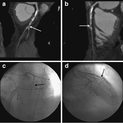
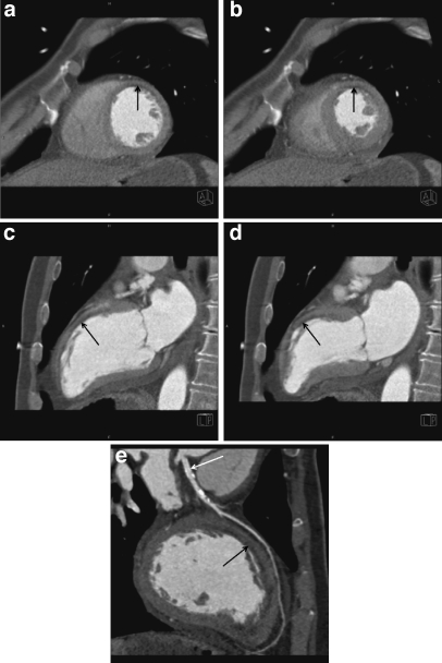
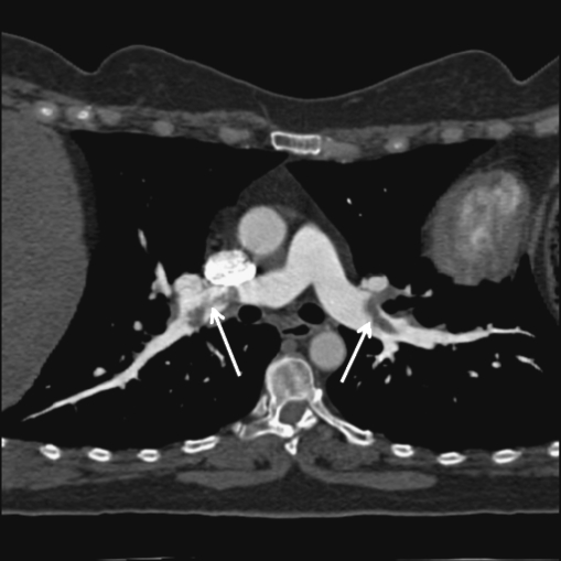
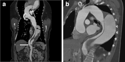
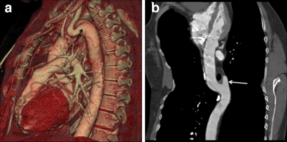
Comprehensive CT angiography protocols offering a simultaneous evaluation of pulmonary embolism, coronary stenoses and aortic disease are gaining attractiveness with recent CT technology. The aim of this study was to assess the diagnostic accuracy of a specific dual-source CT protocol for chest pain assessment. One hundred nine patients suffering from acute chest pain were examined on a dual-source CT scanner with ECG gating at a temporal resolution of 83 ms using a body-weight-adapted contrast material injection regimen. The images were evaluated for the cause of chest pain, and the coronary findings were correlated to invasive coronary angiography in 29 patients (27%). The files of patients with negative CT examinations were reviewed for further diagnoses. Technical limitations were insufficient contrast opacification in six and artifacts from respiration in three patients. The most frequent diagnoses were coronary stenoses, valvular and myocardial disease, pulmonary embolism, aortic aneurysm and dissection. Overall sensitivity for the identification of the cause of chest pain was 98%. Correlation to invasive coronary angiography showed 100% sensitivity and negative predictive value for coronary stenoses. Dual-source CT offers a comprehensive, robust and fast chest pain assessment.


Figures

Fig. 1
Fig. 1
High grade stenosis (arrows) of the left anterior descending coronary artery. a Curved multiplanar reconstruction along the centerline of the vessel. b Orthogonal curved multiplanar reconstruction. c Left anterior oblique-cranial projection at invasive angiography. d Respective right anterior oblique projection
Fig. 2
Fig. 2
Subendocardial scar (arrows) from a myocardial infarction in the territory of the left anterior descending coronary artery. a Diastolic mid-ventricular short axis view. b Systolic short axis view. c Diastolic left two-chamber view. d Systolic two-chamber view. Note the dyskinesia bulging out of the anterior wall. e Multiplanar reconstruction of the left anterior descending coronary artery. Note the stent (white arrow) in the proximal vessel, which had been implanted after interventional re-perfusion of the occluded vessel
Fig. 3
Fig. 3
Curved multiplanar reconstruction of the lower lobe pulmonary arteries showing emboli in the lobar and segmental arteries
Fig. 4
Fig. 4
Dissection of the descending aorta. a Multiplanar reconstruction. Arrows indicate the depiction of the left coronary artery in the gated reconstruction (black arrow), the dissection membrane (white arrow) and the junction between the gated scan and the non-gated scan, which had been continued in this case to include the whole extent of the dissection (outlined arrow). b Angulated para-coronal reconstruction showing the topography
Fig. 5
Fig. 5
Kommerell’s diverticulum (arrows). a Volume-rendered reconstruction showing the origin from the aortic arch and the course of the subclavian artery. Note the median-right course of the descending aorta ventral to the spine. b Curved multiplanar reconstruction showing the proximity to the trachea

References

    1. {'text': '', 'ref_index': 1, 'ids': [{'type': 'DOI', 'value': '10.2214/AJR.05.1153', 'is_inner': False, 'url': 'https://doi.org/10.2214/ajr.05.1153'}, {'type': 'PubMed', 'value': '17179348', 'is_inner': True, 'url': 'https://pubmed.ncbi.nlm.nih.gov/17179348/'}]}
    2. Johnson TRC, Nikolaou K, Wintersperger BJ, Knez A, Boekstegers P, Reiser MF, Becker CR (2007) ECG gated 64 slice CT angiography for the differential diagnosis of acute chest pain. AJR Am J Roentgenol 188:76–82 - PubMed
    1. {'text': '', 'ref_index': 1, 'ids': [{'type': 'DOI', 'value': '10.1007/s11547-006-0044-1', 'is_inner': False, 'url': 'https://doi.org/10.1007/s11547-006-0044-1'}, {'type': 'PubMed', 'value': '16779535', 'is_inner': True, 'url': 'https://pubmed.ncbi.nlm.nih.gov/16779535/'}]}
    2. Savino G, Herzog C, Costello P, Schoepf UJ (2006) 64 slice cardiovascular CT in the emergency department: concepts and first experiences. Radiol Med (Torino) 111:481–496 - PubMed
    1. {'text': '', 'ref_index': 1, 'ids': [{'type': 'PubMed', 'value': '16037533', 'is_inner': True, 'url': 'https://pubmed.ncbi.nlm.nih.gov/16037533/'}]}
    2. White CS, Kuo D, Kelemen M, Jain V, Musk A, Zaidi E, Read K, Sliker C, Prasad R (2005) Chest pain evaluation in the emergency department: can MDCT provide a comprehensive evaluation? AJR Am J Roentgenol 185:533–540 - PubMed
    1. {'text': '', 'ref_index': 1, 'ids': [{'type': 'DOI', 'value': '10.2214/AJR.04.1882', 'is_inner': False, 'url': 'https://doi.org/10.2214/ajr.04.1882'}, {'type': 'PubMed', 'value': '16714608', 'is_inner': True, 'url': 'https://pubmed.ncbi.nlm.nih.gov/16714608/'}]}
    2. Raptopoulos VD, Boiselle PB, Michailidis N, Handwerker J, Sabir A, Edlow JA, Pedrosa I, Kruskal JB (2006) MDCT angiography of acute chest pain: evaluation of ECG-gated and nongated techniques. AJR Am J Roentgenol 186:S346–356 - PubMed
    1. {'text': '', 'ref_index': 1, 'ids': [{'type': 'DOI', 'value': '10.1097/01.rct.0000236421.35761.7a', 'is_inner': False, 'url': 'https://doi.org/10.1097/01.rct.0000236421.35761.7a'}, {'type': 'PubMed', 'value': '17414765', 'is_inner': True, 'url': 'https://pubmed.ncbi.nlm.nih.gov/17414765/'}]}
    2. Johnson TRC, Nikolaou K, Wintersperger BJ, Fink C, Rist C, Leber AW, Knez A, Reiser MF, Becker CR (2007) Optimization of contrast material administration for electrocardiogram-gated computed tomographic angiography of the chest. J Comput Assist Tomogr 31:265–271 - PubMed