. 2007 Dec;37(12):3339-51.
doi: 10.1002/eji.200737752.

Anti-chromatin antibodies drive in vivo antigen-specific activation and somatic hypermutation of rheumatoid factor B cells at extrafollicular sites

Affiliations

Anti-chromatin antibodies drive in vivo antigen-specific activation and somatic hypermutation of rheumatoid factor B cells at extrafollicular sites

Robin A Herlands et al. Eur J Immunol. 2007 Dec.

Abstract

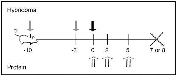
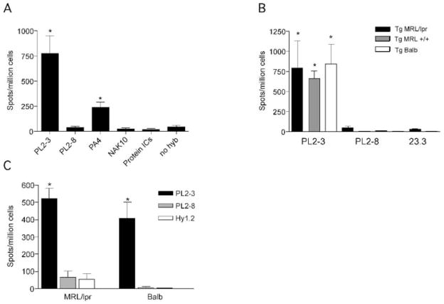
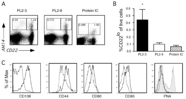
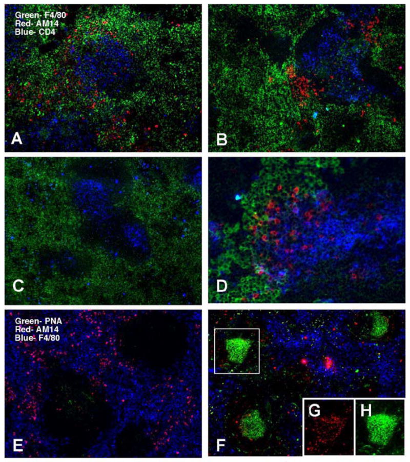
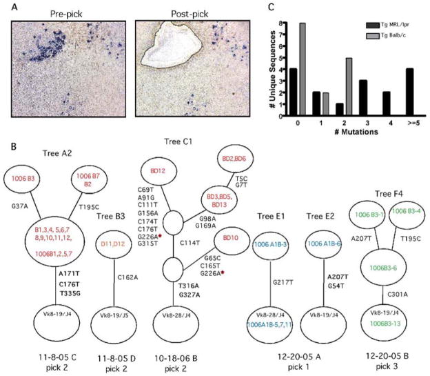
A dominant type of spontaneous autoreactive B cell activation in murine lupus is the extrafollicular generation of plasmablasts. The factors governing such activation have been difficult to identify due to the stochastic onset and chronic nature of the response. Thus, the ability to induce a similar autoreactive B cell response with a known autoantigen in vivo would be a powerful tool in deciphering how autoimmune responses are initiated. We report here the establishment and characterization of a system to initiate autoreactive extrafollicular B cell responses, using IgG anti-chromatin antibodies, that closely mirrors the spontaneous response. We demonstrate that exogenously administered anti-chromatin antibody, presumably by forming immune complexes with released nuclear material, drives activation of rheumatoid factor B cells in AM14 Tg mice. Anti-chromatin elicits autoreactive B cell activation and development into antibody-forming cells at the T zone/red pulp border. Plasmablast generation occurs equally in BALB/c, MRL/+ and MRL/lpr mice, indicating that an autoimmune-prone genetic background is not required for the induced response. Importantly, infused IgG anti-chromatin induces somatic hypermutation in the absence of a GC response, thus proving the extrafollicular somatic hypermutation pathway. This system provides a window on the initiation of an autoantibody response and reveals authentic initiators of it.


Conflict of interest statement

Conflict of interest: The authors declare no conflict of interest.

Figures

Figure 1
Figure 1
Experimental design for the use of IgG2a anti-chromatin antibodies to acutely raise levels of IgG2a RF self-antigen. For hybridoma treatment: mice injected i.p. with pristane on days -10 and -3 (gray arrows), followed by hybridoma injection on Day 0 (black arrow), and sacrifice on Day 7. For protein treatment: mice were injected with 0.5mg purified antibody on Days 0, 2, and 5 (white arrows) and were sacrificed on Day 8.
Figure 2
Figure 2
Anti-nuclear antibodies cause AFC formation and antibody secretion. (A) 4-44+ ELISPOTs from splenocytes of Tg MRL/lpr mice treated with antibody secreting hybridoma or protein ICs. Treatment groups are listed on the x-axis. PL2-3: IgG2a anti-chromatin (n=35); PL2-8: IgG2b anti-chromatin (n=11); PA4: IgG2a anti-DNA (n=4); NAK: IgG2a anti-desmoglein (n=11); protein ICs: ICs of 23.3 (IgG2a anti-NP) and NP-CGG (n=3). No hybridoma: pristane only (n=11). *p<0.05 by Mann-Whitney U test, compared to PL2-8 group (negative control). (B) 4-44+ ELISPOTs from splenocytes of Tg MRL/lpr (black bars), Tg MRL+/+ (gray bars) or Tg BALB/c mice (open bars) treated with PL2-3 (n=5-6 for each strain), PL2-8 or 23.3 hybridoma. *p<0.05 by Mann-Whitney U test, compared to combined PL2-8 data. (C) 4-44+ ELISPOTs from splenocytes of Tg MRL/lpr (n==26, 5, 7 respectively) or Tg BALB/c (n=5, 4, 8 respectively) mice treated with purified PL2-3 (black bars), PL2-8 (gray bars), or Hy1.2 (IgG2a anti-TNP, open bars) for 1 week. *p<0.001 by Mann-Whitney U test, compared to PL2-8 group (negative control).
Figure 3
Figure 3
Anti-chromatin antibodies cause RF B cell plasmablast differentiation as seen by FACS analysis. (A) Representative FACS plots of live spleen cells demonstrating that H Tg MRL/lpr mice treated with PL2-3, but not PL2-8 hybridoma or protein ICs, develop 4-44+/CD22 lo cells, indicative of plasmablast development [26]. (B) Combined data from multiple experiments. PL2-3, n=22; PL2-8, n=12; Protein ICs, n=6. *p=0.0007; ** p=0.0013 by Mann Whitney U test. (C) Histograms of expression of CD138, CD44, CD80 and CD86 gated on live 4-44+ CD22lo (solid line) and 4-44+ CD22hi (dashed line) subsets from mice treated with PL2-3. PNA was used to identify germinal center cells, with 4-44+CD22 hi cells from mice treated with protein ICs serving as a positive control (shaded gray).
Figure 4
Figure 4
AM14 B cells activated by anti-chromatin antibodies localize to the T zone-red pulp border but not GCs in Tg in spleens of MRL/lpr mice. (A–D) AM14 idiotype (4-44) is shown in red, macrophage marker F4/80 in green, and CD4 in blue. (A–C) original magnification 100x; (D) 200x. (A) Tg AM14 MRL/lpr mouse treated for 1 week with PL2-3 hybridoma shows 4-44+ B cells at the T zone-red pulp border. (B) Same as (A) but mice were treated with PL2-3 protein. (C) Tg AM14 MRL/lpr mouse treated for 1 week with PL2-8 hybridoma had few detectable 4-44+ B cells. (D) Higher magnification of spleen of a PL2-3 treated mouse. (E-H) AM14 idiotype (4-44) is shown in red,, F4/80 in blue, and GC marker PNA in green. (E) AM14 MRL/lpr mouse treated with PL2-3 does not show germinal centers. (F) AM14 MRL/lpr mouse treated with protein ICs developed GCs. (G and H) GCs in these mice are positive for AM14 (G) and PNA (H) as shown by the single channel images of the region boxed in (F).
Figure 5
Figure 5
Somatic hypermutation occurs at extrafollicular sites of Tg MRL/lpr and Tg BALB/c mice given IgG2a anti-chromatin. (A) Immunohistochemistry of a representative microdissected region showing AM14 B cells (blue) in a splenic section from a Tg MRL/lpr mouse treated with PL2-3 protein every other day for 2 weeks. Photos are from before and after laser microdissection. Picks typically contained 10–30 AM14+ B cells. Note that image quality is suboptimal due to use of foil-coated slides required for microdissection. (B) Representative genealogical trees from two Tg MRL/lpr mice (trees A2, B3 and C1) and two Tg BALB/c mice (trees E1, E2, and F4) which demonstrate ongoing mutation as reflected by intraclonal diversification. Two additional clones (B2 and F3) from these same picks had no mutation and are thus fully described in the Tables and not shown. Circles at the base of trees represent germline sequences. Non-mutated sequences actually found in a pick are listed in this circle; if there were no germline isolates the circle has no sequence identifiers. Every other open circle, or “node”, represents one or more clones with the same sequence, again as indicated by sequence identifiers. Open circles are implicit nodes required by the genealogy, although no actual sequences representing those nodes were found. Tree “branches” are the lines between nodes. The positions of and types of nucleotide exchanges are listed alongside branches. (C) Histogram of the distribution of the number of mutations per unique sequence for all Tg MRL/lpr and Tg BALB/c picks. The distributions of mutation between MRL/lpr and BALB/c are significantly different (p=0.005, chi-squared test for trend).

Comment in

Similar articles

Cited by

References

    1. Tan EM. Antinuclear antibodies: diagnostic markers for autoimmune diseases and probes for cell biology. Adv Immunol. 1989;44:93–151. - PubMed
    1. Chan OT, Hannum LG, Haberman AM, Madaio MP, Shlomchik MJ. A novel mouse with B cells but lacking serum antibody reveals an antibody-independent role for B cells in murine lupus. J Exp Med. 1999;189:1639–1648. - PMC - PubMed
    1. Shlomchik MJ, Craft J, Mamula MJ. From T to B and back again: positive feedback in systemic autoimmune disease. Nature Reviews Immunology. 2001;1:147–153. - PubMed
    1. Chan O, Shlomchik MJ. A new role for B cells in systemic autoimmunity: B cells promote spontaneous T cell activation in MRL-lpr/lpr mice. J Immunol. 1998;160:51–59. - PubMed
    1. Eisenberg RA, Craven SY, Warren RW, Cohen PL. Stochastic control of anti-Sm autoantibodies in MRL/Mp-lpr/lpr mice. J Clin Invest. 1987;80:691–697. - PMC - PubMed

Publication types

MeSH terms

LinkOut - more resources