. 2007 Nov 24:15:18.
doi: 10.1186/1746-1340-15-18.

A case report of bilateral synovial chondromatosis of the ankle

Affiliations

A case report of bilateral synovial chondromatosis of the ankle

Heather Shearer et al. Chiropr Osteopat. .

Abstract

Background: Synovial chondromatosis is a rare, generally benign condition which affects synovial membranes. It most commonly involves large joints such as the knee, hip, and elbow, but its presence in smaller joints has also been reported. The diagnosis of synovial chondromatosis is commonly made following a thorough history, physical examination, and radiographic examination. Patients may report pain and swelling within a joint which is often aggravated with physical activity.

Case presentation: A rare case of bilateral synovial chondromatosis of the ankle is reviewed. A 26 year-old male presented with chronic bilateral ankle pain. Physical examination suggested and imaging confirmed multiple synovial chondromatoses bilaterally, likely secondary to previous trauma.

Conclusion: The clinical and imaging findings, along with potential differential diagnoses, are described. Since this condition tends to be progressive but self-limiting, indications for surgery depend on the level of symptomatic presentation in addition to the functional demands of the patient. Following a surgical consultation, it was decided that it was not appropriate to pursue surgery at the present time.


Figures

Figure 1
Figure 1
Lateral right ankle radiograph with evidence of calcified loose bodies (arrow) posterior to the talotibial joint. Small loose bodies are also seen anteriorly to the joint (arrow head).
Figure 2
Figure 2
Left lateral ankle view demonstrating multiple calcified loose bodies likely located in both the flexor hallucis and tibialis posterior tendons (arrow). Loose bodies are also present anterior to the talotibial joint (arrow head).
Figure 3
Figure 3
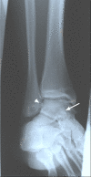
Oblique left ankle radiograph with evidence of calcified loose bodies medial (arrow head) to the lateral maleolus and superimposed over the talus (arrow). This suggests synovial chondromatosis, likely located in both the flexor hallucis and tibialis posterior tendons.
Figure 4
Figure 4
Sagittal MRI of the right ankle (fat-saturated T2-weighted) revealing a predominantly low signal intensity nodule in the synovial sheath of the flexor hallucis longus tendon (arrow).
Figure 5
Figure 5
Axial MRI of the right ankle (proton density) revealing a (arrow) heterogeneous nodule of low and intermediate signal intensities located in the flexor hallucis longus tendon sheath. Of interest is the degree of distension of the tendon sheath secondary to the surrounding effusion.
Figure 6
Figure 6
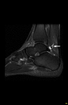
Sagittal MRI of the left ankle (fat-saturated T2-weighted) illustrates (arrow) two distinct low signal intensity nodules with surrounding effusion posterior to the talo-tibial joint.
Figure 7
Figure 7
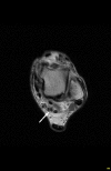
Axial MRI of the left ankle (proton density) demonstrates (arrow) multiple heterogeneous and low signal intensity nodules within the extended flexor hallucis longus tendon sheath.

References

    1. Hocking R, Negrine J. Primary synovial chondromatosis of the subtalar joint affecting two brothers. Foot & Ankle International. 2003;24:865–7. - PubMed
    1. Krebbs VE. The role of hip arthroscopy in the treatment of synovial disorders and loose bodies. Clin Orthop Rel Rsch. 2003;406:48–59. doi: 10.1097/00003086-200301000-00009. - DOI - PubMed
    1. Taconis WK, van der Heul RO, Taminiau AMM. Synovial chondrosarcoma: report of a case and review of the literature. Skeletal Radiol. 1997;26:682–5. doi: 10.1007/s002560050312. - DOI - PubMed
    1. Yu GV, Zema RL, Johnson RWS. Synovial Osteochondromatosis. A case report and review of the literature. J Am Podiatr Med AssocJournal. 2002;92:247–54. - PubMed
    1. Wu CW, Chen YK, Lin LM, Huang E. Primary synovial chondromatosis of the temporomandibular joint. J Otolaryngol. 2004;33:114–9. doi: 10.2310/7070.2004.02111. - DOI - PubMed

LinkOut - more resources