. 2007 Nov 26:5:11.
doi: 10.1186/1479-0556-5-11.

Differential intracellular distribution of DNA complexed with polyethylenimine (PEI) and PEI-polyarginine PTD influences exogenous gene expression within live COS-7 cells

Affiliations

Differential intracellular distribution of DNA complexed with polyethylenimine (PEI) and PEI-polyarginine PTD influences exogenous gene expression within live COS-7 cells

Stephen R Doyle et al. Genet Vaccines Ther. .

Abstract

Background: Polyethylenimine (PEI) is one of the most efficient and versatile non-viral vectors available for gene delivery. Despite many advantages over viral vectors, PEI is still limited by lower transfection efficiency compared to its viral counterparts. Considerable investigation is devoted to the modification of PEI to incorporate virus-like properties to improve its efficacy, including the incorporation of the protein transduction domain (PTD) polyarginine (Arg); itself demonstrated to facilitate membrane translocation of molecular cargo. There is, however, limited understanding of the underlying mechanisms of gene delivery facilitated by both PEI and PEI-bioconjugates such as PEI-polyarginine (PEI-Arg) within live cells, which once elucidated will provide valuable insights into the development of more efficient non-viral gene delivery vectors.

Methods: PEI and PEI-Arg were investigated for their ability to facilitate DNA internalization and gene expression within live COS-7 cells, in terms of the percentage of cells transfected and the relative amount of gene expression per cell. Intracellular trafficking of vectors was investigated using fluorescent microscopy during the first 5 h post transfection. Finally, nocodazole and aphidicolin were used to investigate the role of microtubules and mitosis, respectively, and their impact on PEI and PEI-Arg mediated gene delivery and expression.

Results: PEI-Arg maintained a high cellular DNA uptake efficiency, and facilitated as much as 2-fold more DNA internalization compared to PEI alone. PEI, but not PEI-Arg, displayed microtubule-facilitated trafficking, and was found to accumulate within close proximity to the nucleus. Only PEI facilitated significant gene expression, whereas PEI-Arg conferred negligible expression. Finally, while not exclusively dependent, microtubule trafficking and, to a greater extent, mitotic events significantly contributed to PEI facilitated gene expression.

Conclusion: PEI polyplexes are trafficked by an indirect association with microtubules, following endosomal entrapment. PEI facilitated expression is significantly influenced by a mitotic event, which is increased by microtubule organization center (MTOC)-associated localization of PEI polyplexes. PEI-Arg, although enhancing DNA internalization per cell, did not improve gene expression, highlighting the importance of microtubule trafficking for PEI vectors and the impact of the Arg peptide to intracellular trafficking. This study emphasizes the importance of a holistic approach to investigate the mechanisms of novel gene delivery vectors.


Figures

Figure 1
Figure 1
Efficiency of PEI- and PEI-Arg polyplex internalization. The percentage of fluorescently positive cells (A) and the relative amount of fluorescence per fluorescently positive cell (B) were calculated for polyplexes composed of PEI (■) and PEI-Arg (▲) complexed with YOYO1-labelled pCH110, as detected by FACS 24 h post transfection. Data points represented as mean values ± SEM (N = 3).
Figure 2
Figure 2
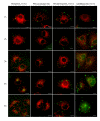
Intracellular trafficking of PEI- and PEI-Arg polyplexes. Time points were taken on the hour, for the first 5 hours post transfection, and viewed by fluorescent microscopy. Merged images were constructed from individual labelled-PEI-, or labelled-pDNA-, and MitoTracker fluorescent images, and are representative of a typical image obtained from five images taken per time point, from two independent experiments. Scale bar = 10 μm.
Figure 3
Figure 3
Effect of microtubule disrupting agent, nocodazole, on microtubule morphology and PEI polyplex trafficking. Untreated (A) and nocodazole treated cells (D) displaying normal and disrupted microtubules, highlighted by anti-β-tubulin visualized using FITC secondary antibody. Nucleus stained with DAPI (B, E) and merged immunofluorescence displayed as (C, F). Distribution of PEI/pDNA-YOYO in untreated (G) and nocodazole treated (J) cells with MitoTracker staining (H, K). Merge MitoTracker and PEI/pDNA-YOYO in untreated (I) and nocodazole treated (L) cells. Scale bar = 10 μm. Images represent typical result of two individual slides viewed for each sample, analyzed 5 h post transfection.
Figure 4
Figure 4
Gene expression efficiency mediated by PEI- and PEI-Arg polyplexes. The percentage of GFP positive cells (A) and the relative amount of fluorescence per GFP positive cell (B) was calculated for polyplexes composed of PEI (■) and PEI-Arg (▲) complexed with pEOTCGFP, as detected by FACS 24 h post transfection. Data represented as mean values ± SEM (N = 3).
Figure 5
Figure 5
Effect of nocodazole and aphidicolin on PEI polyplex mediated GFP expression. Expression of GFP 24 h post transfection in the presence of nocodazole (light bar), or aphidicolin (dark bar), as a percentage of optimal PEI/pDNA mediated GFP expression at N/P 8. Data obtained as detected by FACS 24 h post transfection, and represented as mean values ± SEM (N = 2).

References

    1. Guillem VM, Aliño SF. Transfection pathways of nonspecific and targeted PEI-polyplexes. Gene Therapy and Molecular Biology. 2004;8:369–384.
    1. Lungwitz U, Breunig M, Blunk T, Gopferich A. Polyethylenimine-based non-viral gene delivery systems. European Journal of Pharmaceutics & Biopharmaceutics. 2005;60:247–266. doi: 10.1016/j.ejpb.2004.11.011. - DOI - PubMed
    1. Florea BI, Meaney C, Junginger HE, Borchard G. Transfection efficiency and toxicity of polyethylenimine in differentiated Calu-3 and nondifferentiated COS-1 cell cultures. AAPS PharmSci. 2002;4:E12. doi: 10.1208/ps040312. - DOI - PMC - PubMed
    1. Zaric V, Weltin D, Erbacher P, Remy JS, Behr JP, Stephan D. Effective polyethylenimine-mediated gene transfer into human endothelial cells. J Gene Med. 2004;6:176–184. doi: 10.1002/jgm.484. - DOI - PubMed
    1. Boussif O, Lezoualc'h F, Zanta MA, Mergny MD, Scherman D, Demeneix B, Behr JP. A versatile vector for gene and oligonucleotide transfer into cells in culture and in vivo: polyethylenimine. Proc Natl Acad Sci USA. 1995;92:7297–7301. doi: 10.1073/pnas.92.16.7297. - DOI - PMC - PubMed

LinkOut - more resources