. 2008 Jan;134(1):292-305.
doi: 10.1053/j.gastro.2007.10.030. Epub 2007 Oct 25.

Disordered pancreatic inflammatory responses and inhibition of fibrosis in CD39-null mice

Affiliations

Disordered pancreatic inflammatory responses and inhibition of fibrosis in CD39-null mice

Beat M Künzli et al. Gastroenterology. 2008 Jan.

Abstract

Background & aims: Extracellular nucleotides are released from injured cells and bind purinergic-type 2 receptors (P2-Rs) that modulate inflammatory responses. Ectonucleotidases, such as CD39/nucleoside triphosphate diphosphohydrolase-1, hydrolyze extracellular nucleotides to integrate purinergic signaling responses. Because the role of extracellular nucleotides and CD39 in mediating inflammation and fibrosis are understood poorly, we studied the impact of CD39 gene deletion in a model of pancreatic disease.

Methods: Pancreatitis was induced by cyclosporine pretreatment, followed by cerulein injections (50 mug/kg, 6 intraperitoneal injections/day, 3 times/wk); mice were killed at day 2, week 3, and week 6. Experimental parameters were correlated with cytokine levels in blood, RNA, and protein expression of purinergic and fibrosis markers in tissues. Immunohistochemistry and pancreatic morphometry of fibrosis were performed in wild-type and CD39-null mice. Effects of CD39 deletion on proliferation of primary pancreatic stellate cells (PSCs) were investigated in vitro.

Results: Wild-type mice developed morphologic features of pancreatitis with the anticipated development of parenchymal atrophy and fibrosis. CD39 and P2-R became overexpressed in vascular and adventitious wild-type tissues. In contrast, CD39-null mice had inflammatory reactions but developed only minor pancreatic atrophy and limited fibrosis. Interferon-gamma became significantly increased in tissues and plasma of CD39-null mice. Wild-type PSCs expressed high levels of CD39 and P2-R. CD39-null PSCs showed decreased rates of proliferation and the expression of procollagen-alpha1 was inhibited significantly in vitro (P < .03).

Conclusions: CD39 deletion decreases fibrogenesis in experimental pancreatitis. Our data implicate extracellular nucleotides as modulators of PSC proliferation and collagen production in pancreatitis.


Figures
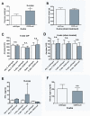
Figure 1
Figure 1
Assessment of pancreatitis. (A) After 6 wks of CIP, significant decreases in pancreas weights of wt versus CD39-null mice were observed (secondary to atrophy of the pancreas, P<0.005). (B) Saline injection for 6 wks did not affect pancreas weights (sham treatment). (C, D) Blood amylase activity revealed no differences between cerulein- or sham treated wt and CD39-null mice (normal range of used test, up to 100 IU/L). (E) Heightened levels of IFN-γ were only detected in sera from CD39-null mice after 3 wks of pancreatitis induction (P<0.05). (F) Blood sugar levels remained normal and did not differ between the treatment groups.
Figure 2
Figure 2
Western analyses. Protein expression was determined from baseline pancreas without treatment, after 3 wks and 6 wks of CIP. (A) Representative samples showing high induction of CD39 at 3 and 6 wks of CIP in wt mice. CD39-null mice show increased protein expression of CD39L1 and P2X7 (at 3 wks). P2Y2 was upregulated after 6 wks. MMP-2 was significantly upregulated but only in CD39-null mice. TGF-β1 was equally upregulated at 3 wks of CIP. CD45 was predominantly expressed in CD39-null mice at 3 wks of CIP treatment. (A) Fibronectin was highly upregulated at 6 wks treatment, more prominently expressed in wt mice. Each lane (40 μg total protein/lane) represents one pancreas sample. Shown is one representative individual mouse per group (baseline pancreas, CIP at 3 and 6 wks time, respectively). (B) Protein lysates from primary cultured PSC showed high expression for CD39. P2X7 expression was more prominent in wt cells when compared to CD39-null PSC. Protein levels of fibronectin were elevated in wt PSC versus CD39-null PSC.
Figure 3
Figure 3
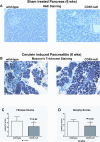
Pancreas morphology. (A) Pancreas architecture in sham treated remained unchanged in wt and CD39-null mice. (B) Panels show representative Masson's trichrome staining sections from wt and CD39-null mice. Histological parameters such as fibrosis (C) (P<0.02) and atrophy (D) (P<0.02) revealed significant differences between wt and CD39-null pancreases.
Figure 4
Figure 4
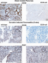
Immunohistochemistry in cerulein induced pancreatitis. (A, B) Panels show representative immunohistochemical analyses of CD39 and P2X7 in tissues of wt and CD39-null mice after 6 wks of CIP. Normal pancreas of sham treated wt and CD39-null mice did not reveal any signs of significant architectural changes after saline treatment. (B, D) There was no reactivity for CD39 in CD39-null mice. During pancreatitis, the density of CD39 positive staining increased and localized to endothelial and cells in the interacinar and interseptal space (insert Figure 3C), that share homologies with myofibroblast like cells and resembling PSC in localization and morphology. (E, F) P2X7 is mainly localized to immune cells (LC = leukocyte) and elements of the vasculature.
Figure 5
Figure 5
Immunohistochemistry and immunofluorescence in chronic pancreatitis. (A, B) CD34, a myofibroblast- and stromal cell marker localized within the periacinar and periductular areas. CD39 is colocalized with CD34. (C, D) The double positive cells display a distinct morphology, are single cells with spindle like protrusions and comparable with the described phenotype and morphology of PSC. (E, F) CD39L1 was colocalized to CD34 in the rarely encountered areas of dense fibrosis in CD39-null mice (insert Figure 4F), whereas in dense fibrotic areas of wt mice, co-localization was only minimal.
Figure 6
Figure 6
Phenotype and differentiation of PSC in primary cultures. (A, B) CD39-null PSC displayed a highly significant proliferation deficit in primary culture compared to wt PSC (P<0.02). (A) Stimulation of CD39-null PSC with PDGF (5 ng/ml) did not lead to increased proliferation in CD39-null PSC, whereas wt PSC responded to PDGF. (C) Elevated concentration of ATP levels (10 and 50 μmol ATP but not 150 and 400 μmol) in culture media significantly increased proliferation of wt PSC (P<0.05). (D) Significant differences in mRNA expression profile for CD39 and P2X7 in cultures of wt PSC compared to CD39-null PSC. CD39-null PSC were viable and differentiated in a comparable manner to wt PSC, as indicated by equal expression of α-SMA on mRNA (E) and morphologically (F). Morphology of PSC revealed high positive staining for α-SMA and desmin in cultured PSC. (F) Differentiation and viability were comparable in wt and CD39-null PSC.
Figure 7
Figure 7
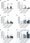
Proliferation defect in CD39-null PSC. In general, a lack of plasticity was noted in CD39-null PSC responses to growth factors and TGF-β. Wt and CD39-null PSC were stimulated with PDGF (5ng/ml) or TGF-β1 (2ng/ml), (A) The mRNA expression profiles reached moderate levels of CD39 expression in wt PSC. (B) CD39L1 levels of expression in wt (and to a lesser extent in CD39-null PSC) decreased when stimulated with PDGF or TGF-β1. P2X7 expression in wt PSC was significantly increased when compared to CD39-null PSC (P<0.03). (C) P2X7 levels decreased during stimulation with PDGF and TGF-β1. (D) Levels of P2Y2 were comparable during stimulation experiments. (E) At baseline conditions and under stimulation with PDGF, α-SMA remained expressed at comparable levels. Procollagen-α1 production following PDGF stimulation was significantly decreased in CD39-null PSC. (F) Most strikingly, procollagen-α1 expression in wt PSC significantly increased when stimulated with the profibrogenic cytokine TGF-β1 (P<0.03). This defect suggests non-responsiveness of and probable defect in collagen production in the CD39-null PSC.

References

    1. Etemad B, Whitcomb DC. Chronic pancreatitis: diagnosis, classification, and new genetic developments. Gastroenterology. 2001;120:682–707. - PubMed
    1. Luttenberger T, Schmid-Kotsas A, Menke A, Siech M, Beger H, Adler G, Grunert A, Bachem MG. Platelet-derived growth factors stimulate proliferation and extracellular matrix synthesis of pancreatic stellate cells: implications in pathogenesis of pancreas fibrosis. Lab Invest. 2000;80:47–55. - PubMed
    1. Apte MV, Phillips PA, Fahmy RG, Darby SJ, Rodgers SC, McCaughan GW, Korsten MA, Pirola RC, Naidoo D, Wilson JS. Does alcohol directly stimulate pancreatic fibrogenesis? Studies with rat pancreatic stellate cells. Gastroenterology. 2000;118:780–94. - PubMed
    1. Bachem MG, Schneider E, Gross H, Weidenbach H, Schmid RM, Menke A, Siech M, Beger H, Grunert A, Adler G. Identification, culture, and characterization of pancreatic stellate cells in rats and humans. Gastroenterology. 1998;115:421–32. - PubMed
    1. Apte MV, Haber PS, Darby SJ, Rodgers SC, McCaughan GW, Korsten MA, Pirola RC, Wilson JS. Pancreatic stellate cells are activated by proinflammatory cytokines: implications for pancreatic fibrogenesis. Gut. 1999;44:534–41. - PMC - PubMed

Publication types