Review
. 2008 Jan 9;582(1):117-31.
doi: 10.1016/j.febslet.2007.11.051. Epub 2007 Nov 26.

TNF-alpha and adipocyte biology

Affiliations
Review

TNF-alpha and adipocyte biology

William P Cawthorn et al. FEBS Lett. .

Abstract

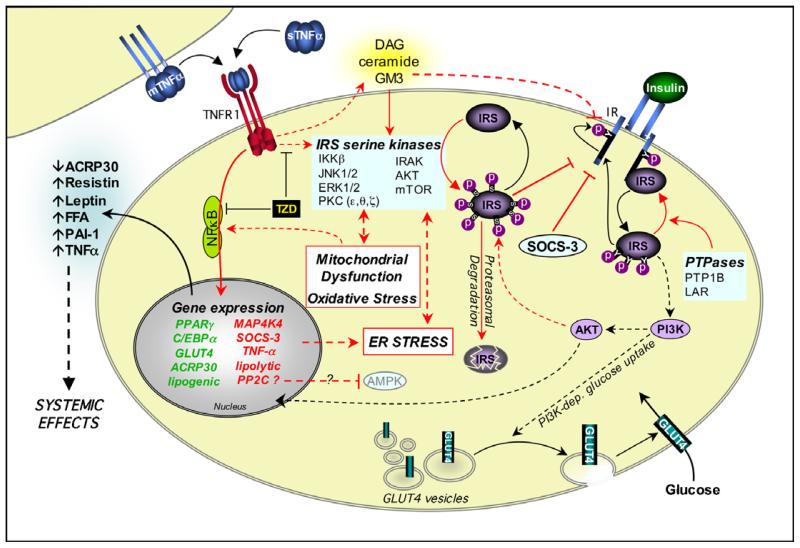
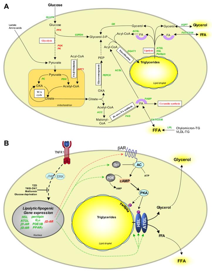
Dyslipidemia and insulin resistance are commonly associated with catabolic or lipodystrophic conditions (such as cancer and sepsis) and with pathological states of nutritional overload (such as obesity-related type 2 diabetes). Two common features of these metabolic disorders are adipose tissue dysfunction and elevated levels of tumour necrosis factor-alpha (TNF-alpha). Herein, we review the multiple actions of this pro-inflammatory adipokine on adipose tissue biology. These include inhibition of carbohydrate metabolism, lipogenesis, adipogenesis and thermogenesis and stimulation of lipolysis. TNF-alpha can also impact the endocrine functions of adipose tissue. Taken together, TNF-alpha contributes to metabolic dysregulation by impairing both adipose tissue function and its ability to store excess fuel. The molecular mechanisms that underlie these actions are discussed.


Figures

Fig. 1
Fig. 1. Signalling pathways induced by TNF-α in adipose tissue
Upon TNFR1 activation the adapter molecule TRADD (TNFR-associated death domain protein) interacts with the TNFR1-DD. TRADD recruits downstream adapter molecules such as Fas-associated death domain protein (FADD), TNFR-associated factor 2 (TRAF2), receptor-interacting protein 1 (RIP1) and mitogen-activated protein kinase (MAPK)-activating death domain protein (MADD). These adapters then mediate the activation of divergent downstream signalling pathways. Apoptosis: FADD recruits caspases such as caspase 8 to TNFR1, thereby forming the TNFR1 death-inducing signalling complex (DISC). This complex activates downstream caspases, leading to apoptosis. Ceramide production: the TNFR1-DD activates acidic sphingomyelinase (aSMase), which hydrolyses sphingolipids to produce ceramide. Ceramides can be converted into toxic lipids, such as the ganglioside GM3. Ceramides and gangliosides may then mediate transcriptional effects, possibly by activation of NFκB. Ceramides can also stimulate apoptosis and impair insulin signalling, and both ceramide and caspases may inhibit AKT. NFκB activation: TRAF2 and RIP1 mediate NFκB activation by recruiting the IKK (inhibitor of NFκB (IκB) kinase) complex to TNFR1. TRAF2 interacts with IKKβ and IKKα, and also promotes the K63-linked polyubiquitination of RIP1 (Ub, ubiquitin); Ubiquitinated RIP1 binds to IKKγ. TRAF2 also recruits TAK1 (TGFβ-activated kinase 1) to TNFR1 via an interaction with TAB1 (TAK1 binding protein 1), which may result in activation of TAK1. This also brings TAK1 into close proximity of the IKK complex. TAK1 may activate IKKβ either by phosphorylating it directly, or by activating NIK (NFκB-inducing kinase), which then phosphorylates IKKβ. Active IKKβ phosphorylates IκBα on conserved serines, leading to its polyubiquitination and proteasomal degradation. This liberates the bound NFκB dimers (e.g. the prototypical p65/p50 heterodimer), which translocate into the nucleus to mediate transcriptional effects. MAPK activation: TNF-α activates various MAPKs in adipocytes, including ERK1/2, p38 MAPK and JNK, possibly via the adapter MADD. ERK1/2 and JNK can each regulate transcription by suppressing the activity of PPARγ. JNK may also exert transcriptional effects via c-jun. JNK, ERK1/2, IKKβ and other kinases have also been implicated in TNF-α-induced serine phosphorylation of IRS-1, which suppresses insulin signalling. Other effects: TNF-α can activate PKA (protein kinase A) by increasing cAMP levels, possibly via transcriptional effects. TNF-α can activate the transcription factor NFAT via Ca2+, however this has not been established in adipocytes. In preadipocytes TNF-α can stimulate transcription by TCF7L2 (transcription factor 7-like 2). The transcriptional effects of TNF-α promote ER stress, oxidative stress and mitochondrial dysfunction, lipolysis and altered adipokine expression, thereby compromising insulin signalling and adipocyte lipid metabolism. TNFR2 may also mediate some effects of TNF-α in adipose tissue, albeit by unknown mechanisms. Factors that have not been directly established in TNF-α-induced signals in adipocytes are faded. Dashed lines indicate effects that are mediated by indirect or unestablished mechanisms. Question marks indicate effects that remain to be fully established. Genes written in red or green are upregulated or downregulated by TNF-α, respectively.
Fig. 2
Fig. 2. Mechanisms of TNF-α-induced insulin resistance in adipose tissue
Insulin mediates metabolic effects by binding to the insulin receptor (IR). The IR has intrinsic tyrosine kinase activity, and insulin binding promotes autophosphorylation of the IR on key intracellular tyrosines. This allows the recruitment of insulin receptor substrates (IRS) such as IRS-1. IR-bound IRS proteins are themselves phosphorylated on tyrosine residues, allowing the activation of signalling pathways such as phosphatidylinositol 3-kinase (PI3K) and AKT. These exert downstream effects such as translocation of GLUT4 from intracellular storage vesicles to the plasma membrane. Protein tyrosine phosphatases (PTPases) can abrogate IR-proximal signalling. TNF-α also impairs IR-proximal signalling through the activation of IRS serine kinases and the production of toxic lipids, as described in Figure 1. TNF-α further compromises insulin action through transcriptional effects, possibly mediated via NFκB activation. These cause downregulation of components required for insulin responsiveness and upregulation of factors that may further impair both local and systemic insulin sensitivity, such as SOCS-3. Transcriptional effects may also promote cellular stresses that can impair insulin signalling. Thiazolidinediones (TZD) impair TNF-α-induced insulin resistance. Black arrows indicate effects that promote insulin signalling. Red arrows indicate effects that antagonise insulin signalling. Effects on gene expression are indicated as described in Fig. 1.
Fig. 3
Fig. 3. Effects of TNF-α on adipocyte lipid metabolism and lipolysis
(A) Pathways involved in adipocyte lipogenesis and their modulation by TNF-α. Adipocytes take up glucose, glycerol and FFA from serum and convert these to triglycerides via numerous biochemical pathways. White boxes indicate the nature of the pathways involved. TNF-α modulates the expression of many of the enzymes and other proteins that regulate these pathways, thereby compromising adipocyte triglyceride storage. (B) Mechanisms of TNF-α-induced lipolysis in adipocytes. During lipolysis triglycerides are hydrolysed into FFA and glycerol. Stimulus-induced lipolysis is mediated by hormone-sensitive lipase (HSL), whereas basal lipolysis may be regulated by adipose triglyceride lipase (ATGL). Lipolysis is further regulated by the perilipins, a family of phosphoproteins that are localised at the surface of lipid droplets in adipocytes. Perilipins normally inhibit lipolysis by preventing access of lipases to the lipid droplets. PKA stimulates lipolysis by phosphorylating HSL and the perilipins. This activates HSL and enables it to access substrates in the lipid droplets. Downregulation of cAMP by phosphodiesterases (PDE) or stimulation of Giα-coupled receptors abrogates PKA activation and thereby inhibits lipolysis. TNF-α stimulates lipolysis via a glucose-dependent mechanism that likely involves transcriptional effects. These may be mediated via JNK, ERK1/2 and NFκB, resulting in upregulation of cAMP and downregulation of perilipins. Some of these effects, such as downregulation of Giα subtypes, are specific to rodent adipocytes. Arrows and gene names in red and green indicate upregulation and downregulation, respectively. PEPCK, phosphoenolpyruvate carboxykinase; ACC, acetyl-CoA carboxylase; FAS, fatty acid synthase; FABP, fatty acid-binding protein; FAT, fatty acid translocase; ACS, aceyl-CoA synthetase long chain; DGAT1, diacylglycerol acyltransferase 1; AQP7, aquaporin 7; GK, glycerol kinase; G3PDH, glyceraldehyde-3-phosphate dehydrogenase; PFK, phosphofructokinase; PGK, phosphoglycerate kinase; PK, pyruvate kinase; PC, pyruvate carboxylase; PDH, pyruvate dehydrogenase; TCA, tri-carboxylic acid; CPT1 carnitine palmitoyltransferase 1; OXA, oxaloacetate; PEP, phosphoenol pyruvate; TG, triglyceride; VLDL, very low-density lipoprotein.

References

    1. Kriegler M, Perez C, DeFay K, Albert I, Lu SD. A novel form of TNF/cachectin is a cell surface cytotoxic transmembrane protein: ramifications for the complex physiology of TNF. Cell. 1988;53:45–53. - PubMed
    1. Black RA, et al. A metalloproteinase disintegrin that releases tumour-necrosis factor-alpha from cells. Nature. 1997;385:729–733. - PubMed
    1. Perez C, Albert I, DeFay K, Zachariades N, Gooding L, Kriegler M. A nonsecretable cell surface mutant of tumor necrosis factor (TNF) kills by cell-to-cell contact. Cell. 1990;63:251–258. - PubMed
    1. Xu H, Sethi JK, Hotamisligil GS. Transmembrane tumor necrosis factor (TNF)-alpha inhibits adipocyte differentiation by selectively activating TNF receptor 1. J. Biol. Chem. 1999;274:26287–26295. - PubMed
    1. Grell M. Tumor necrosis factor (TNF) receptors in cellular signaling of soluble and membrane-expressed TNF. J. Inflamm. 1995;47:8–17. - PubMed

Publication types

Substances